

Clinical features and outcomes of patients admitted to the intensive care unit due to acute abdomen are important to be investigated.
To evaluate the outcomes of critically ill subjects with acute abdomen according to etiology, comorbidity and severity.
Outcomes of 1,523 patients (878 women, mean age 66±18 years) consecutively admitted to a specialized gastrointestinal intensive care unit with different causes of acute abdomen from January 2012 to December 2019, were retrospectively evaluated according to etiology, comorbidity and severity.
The most common causes of acute abdomen were obstructive and inflammatory, particularly large bowel obstruction (27%), small bowel obstruction (18%) and acute pancreatitis (17%). Overall mortality was 13%. Surgery was required in 34% of patients. Median length of stay in the hospital was 9 [1-101] days. On univariate analysis mortality was significantly associated with age, APACHE II, Charlson comorbidity index, requirement for surgery and malignancy (p<0.0001), but only APACHE II, Charlson comorbidity index and surgical interventional remained significant on multivariate analysis.
Critically ill patients admitted to the intensive care unit with acute abdomen constitute a heterogeneous group of subjects with different prognosis. Mortality is more related to the severity of the disease, comorbidity and need for surgery than to the etiology of the acute abdomen.
Cancer patients present various physiological, metabolic, social and emotional changes as a consequence of the disease’s own catabolism, and may be potentiated in the gastrointestinal tract cancer by their interference with food intake, digestion and absorption.
o evaluate the functionality of upper gastrointestinal cancer patients which have undertaken surgery and analyze the factors associated with changes in strength and functionality during hospitalization time.
Prospective analytical study in patients with cancer of the upper gastrointestinal tract which have undertaken surgery. Was evaluated the handgrip strength using a hand dynamometer and functionality through the functional independence measure and Functional Status Scale for Intensive Care Unit in the preoperative period, 2nd and 7th postoperative day.
Were included 12 patients, 75% men, and mean age was 58.17 years old. The most prevalent tumor site was stomach (66.7%). There was a progressive reduction from the pre-operative palmar grip strength to the 2nd and 7th postoperative day, respectively. There was a decrease in functional performance from the preoperative period to the 2nd and a gain from the 2nd to the 7th postoperative day (p<0.001).
An important reduction in the handgrip strength and functionality was evidenced during the postoperative period in relation to the basal value in the pre-operative period.
Collis gastroplasty for esophageal lengthening is a complex adjunct to hiatal hernia repair in patients with esophageal foreshortening.
To study the final morphology of the repair using state-of-the-art imaging: computed tomography with three-dimensional reconstruction.
Nine patients with prior Collis gastroplasty and hiatal hernia repair were studied with three-dimensional computed tomography reconstruction to evaluate the anatomy of the repair and screen for hiatal hernia recurrence. Secondary outcomes were quality of life and surgical morbidity.
After a medium follow-up of 34 months, objective recurrence of the hiatal hernia was observed in three patients (1.5, 2.2, and 3 cm), and two patients were symptomatic. The gastroesophageal junction tube (neo-esophagus) created by the gastroplasty was similar in shape and volume to the native esophagus in all patients. The fundoplication previously performed covered the neo-esophagus in only two of the nine patients. No fistulas or mortality were observed.
Collis gastroplasty reliably produces a tubular gastric conduit resembling the native esophagus in shape and volume. 3D CT is a promising tool for evaluating the postoperative morphology of the repair and anatomical recurrences, demonstrating the durable anatomical structure of Collis gastroplasty.
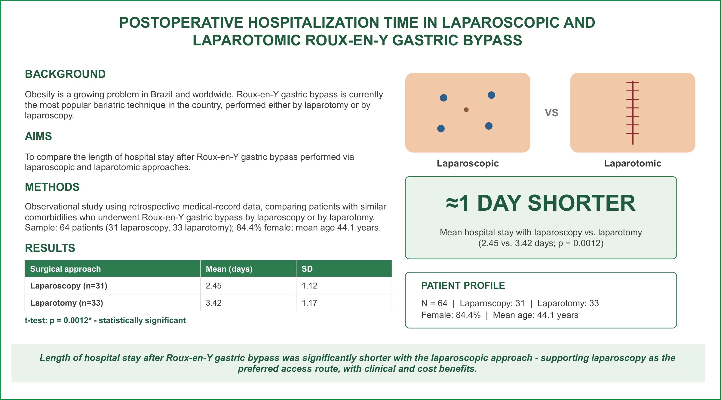
Obesity is a growing problem both in Brazil and worldwide, and bariatric surgery is a well-established method for reducing weight and, consequently, reducing the risk of death attributed to cardiovascular and metabolic problems. Roux-en-Y gastric bypass is currently the most popular surgical technique in Brazil, performed via either laparotomy or laparoscopy.
To compare the length of hospital stay after Roux-en-Y gastric bypass performed via laparoscopic and laparotomic approaches.
An observational study using retrospective data from medical records, comparing patients who underwent Roux-en-Y gastric bypass by laparoscopy and patients operated on by laparotomy, with similar comorbidities.
The sample consisted of 64 patients who underwent surgery: 31 via laparoscopy and 33 via laparotomy, of whom 84.4% were female, with a mean age of 44.1 (SD=10.1). 15.6% were male, with a mean age of 40.2 (SD=13.3). Among the four variables evaluated (sex, age, length of hospital stay, and type of surgery), the only statistically significant difference found was in the length of hospital stay according to each type of surgery, with patients undergoing Roux-en-Y gastroplasty via laparoscopic approach remaining hospitalized for a mean of 2.45 days (SD=1.12), while those undergoing the same surgery via laparotomic approach remained hospitalized for a mean of 3.42 days (SD=1.17).
The findings of this study indicate a numerically and statistically significant difference in length of hospital stay between both surgical access routes for bariatric surgery, with a shorter stay observed in the laparoscopic approach. This information is relevant, as it has financial impact by reducing bed occupancy time and the costs arising from hospitalization.
Among the 2.0 billion overweight individuals in the world, Brazil ranks fifth in the number of obese people, therefore requiring treatment options for obesity.
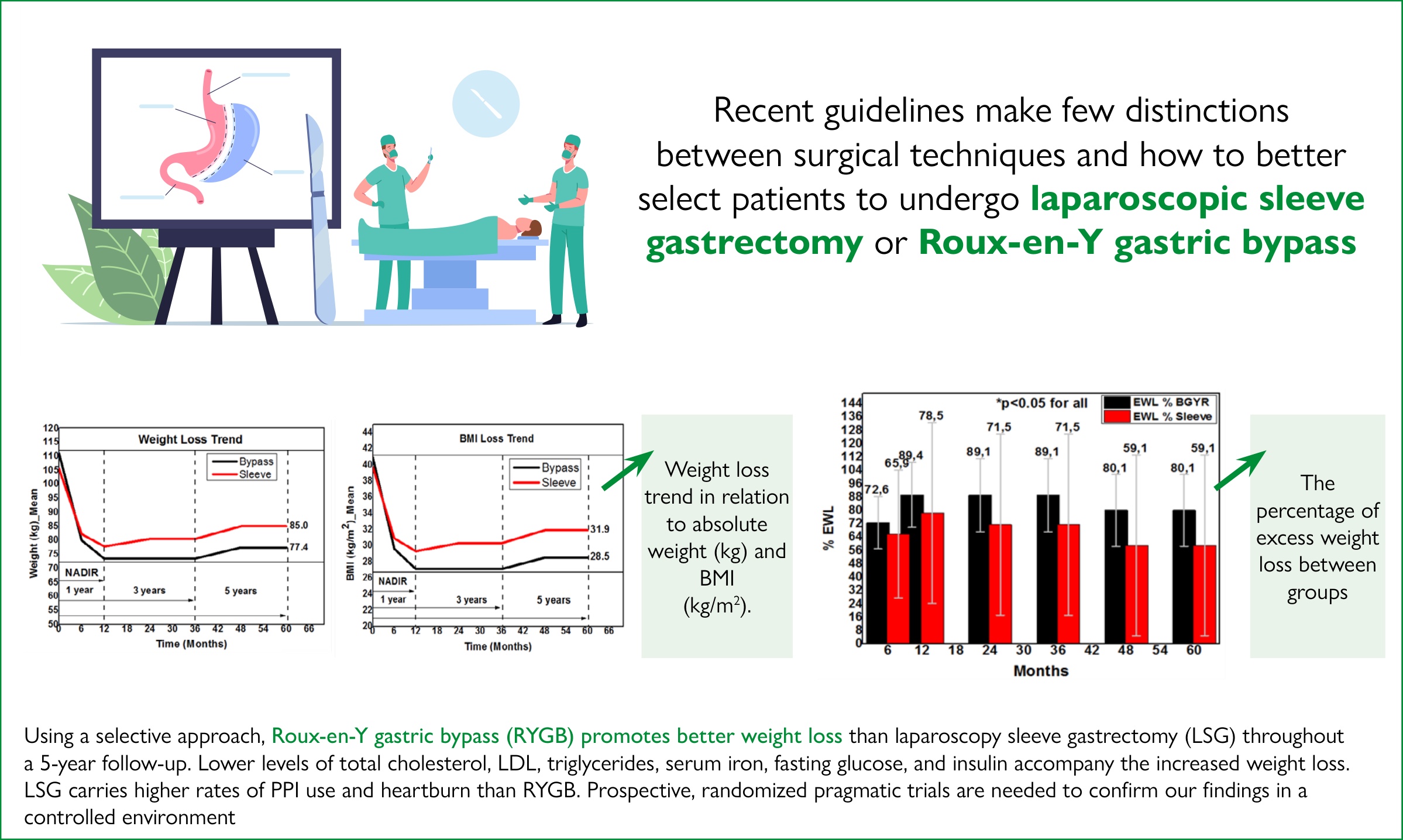
The aim of this study was to compare the percentage of total body weight loss (%TWL), change in body mass index (BMI), percentage of excess weight loss (%EWL), incidence of reflux esophagitis, and occurrence of Barrett's esophagus in obese patients undergoing gastric bypass (Roux-en-Y gastric bypass [RYGB]) and sleeve gastrectomy (SG), both techniques by videolaparoscopy.
The study included 100 consecutive patients who underwent RYGB and SG techniques, totaling 200 patients, and were followed up for 60 months, from June 2013 to July 2018.
The frequency of gastroesophageal reflux disease (GERD) was lower in RYGB patients (p<0.05). At 60 months, the %EWL was 77.4±13.3 kg (RYGB) versus 80.5±17.5 kg (SG) (p<0.05). The BMI data were statistically significantly different between groups after 5 years (28.5±3.9 kg/m2 in RYGB and 31.9±5.3 kg/m2 in SG groups, p<0.05). During the follow-up, the RYGB showed higher %EWL compared to the SG (at 60 months, 80.1% vs. 59.1%, respectively, p<0.05). The %TWL was 30% for the RYGB and 19.7% for the SG (p<0.05). The RYGB had a lower frequency of reflux esophagitis and Barrett's esophagus.
The RYGB technique showed greater absolute weight loss, %TWL, BMI reduction, and %EWL in higher obesity classes compared to the SG technique. Additionally, RYGB had a lower frequency of reflux esophagitis and Barrett's esophagus.
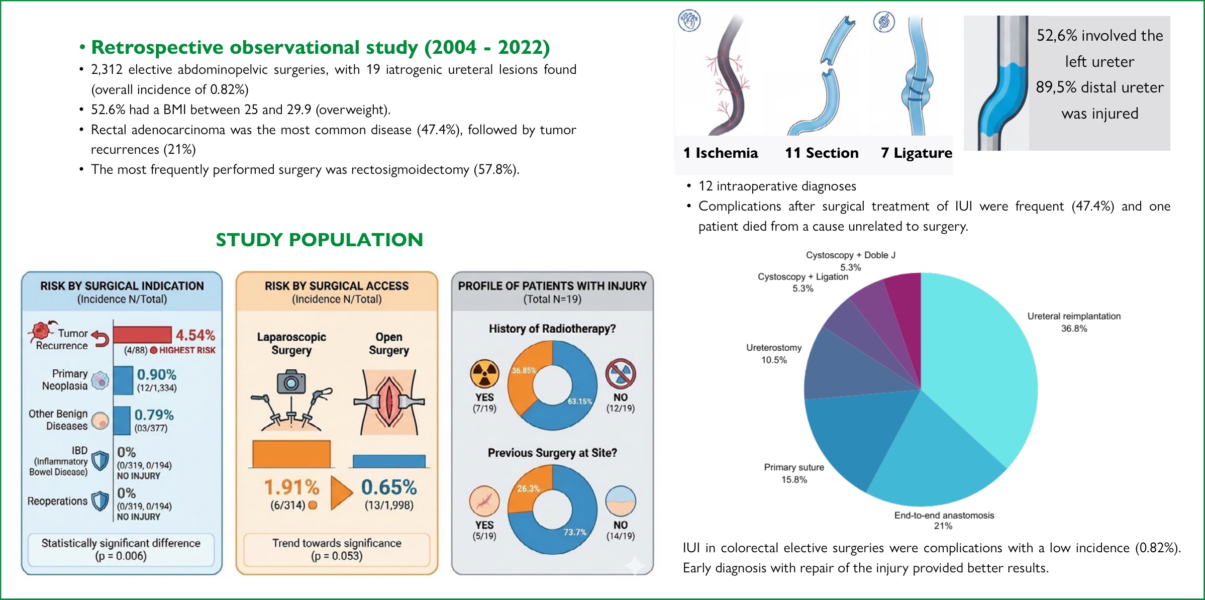
Iatrogenic ureteral injuries (IUI) are rare surgical complications in abdominopelvic surgeries, with an incidence varying between 0.15 and 1.0%.
To evaluate the incidence of IUI in elective colorectal surgeries performed in a tertiary university hospital.
This is a retrospective analysis of patients operated from 2004 to 2022, who presented IUI. Demographic data, underlying disease, predisposing factors, surgery access, location of the lesions and their characteristics, diagnosis time, treatment carried out, and follow-up were analyzed.
In the period, 2,312 abdominopelvic surgeries were performed, of which 1,998 were open and 314 were laparoscopic, with 19 IUI (0.82%). The mean age was 55.6 years, 57.9% were male, and 89.5% were white. The majority of patients were overweight (52.6%), and 73.7% had a history of abdominal surgery. Primary rectal adenocarcinoma was the most common disease (47.4%), followed by tumor recurrences (21.0%). IUI occurred in 1.91% of laparoscopic surgeries and 0.65% of open surgeries (p=0.053); patients with tumor recurrence presented more IUI than those with primary tumors or benign diseases (p=0.006). They were commonly observed in the left ureter (52.6%) and in the distal portion (89.5%), the main mechanism being the section (57.9%). Intraoperative diagnosis occurred in 12 patients (63.2%). IUI correction was predominant in ureteral reimplantation and end-to-end ureteral anastomosis. Postoperative complications were common (47.4%), and one patient died from causes unrelated to surgery.
IUI presented low incidence in colorectal elective surgeries and were more frequent in surgeries for tumor recurrences, in the left ureter and the distal third. Early diagnosis with repair of the injury provided better results.
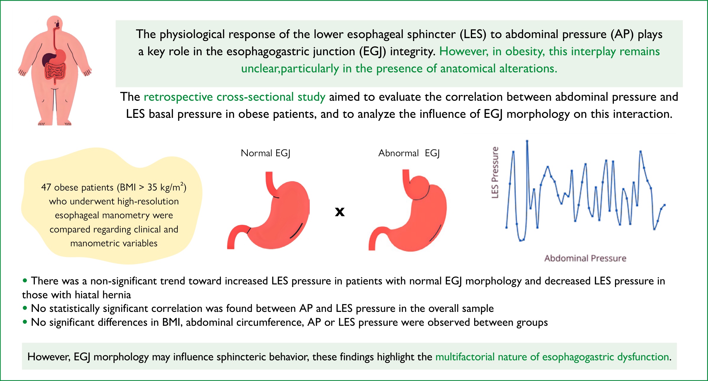
The physiological response of the lower esophageal sphincter (LES) to abdominal pressure (AP) plays a key role in the esophagogastric junction (EGJ) integrity. However, in obesity, this interplay remains unclear, particularly in the presence of anatomical alterations.
This study aims to evaluate the correlation between abdominal pressure and LES basal pressure in obese patients, and to analyze the influence of EGJ morphology on this interaction.
This retrospective cross-sectional study included 47 obese patients (BMI>35 kg/m2) who underwent high-resolution esophageal manometry. Patients were divided into two groups based on EGJ morphology: normal (Type I) and abnormal (Types II/III, hiatal hernia) and were compared based on clinical and manometric variables. Correlation analyses between AP and LES pressure were performed for the entire sample and stratified by EGJ morphology.
No statistically significant correlation was found between AP and LES pressure in the overall sample (rho=0.05; p=0.737). In the subgroup analysis, there was a non-significant trend toward increased LES pressure in patients with normal EGJ morphology (r=0.26; p=0.150) and decreased LES pressure in those with hiatal hernia (r=-0.39; p=0.131). No significant differences in BMI, abdominal circumference, AP, or LES pressure were observed between groups.
In obese individuals, abdominal pressure does not show correlation with LES pressure. However, EGJ morphology may influence sphincteric behavior, with a trend toward LES pressure increase in normal EGJ and reduction in altered morphologies. These findings highlight the multifactorial nature of esophagogastric dysfunction.
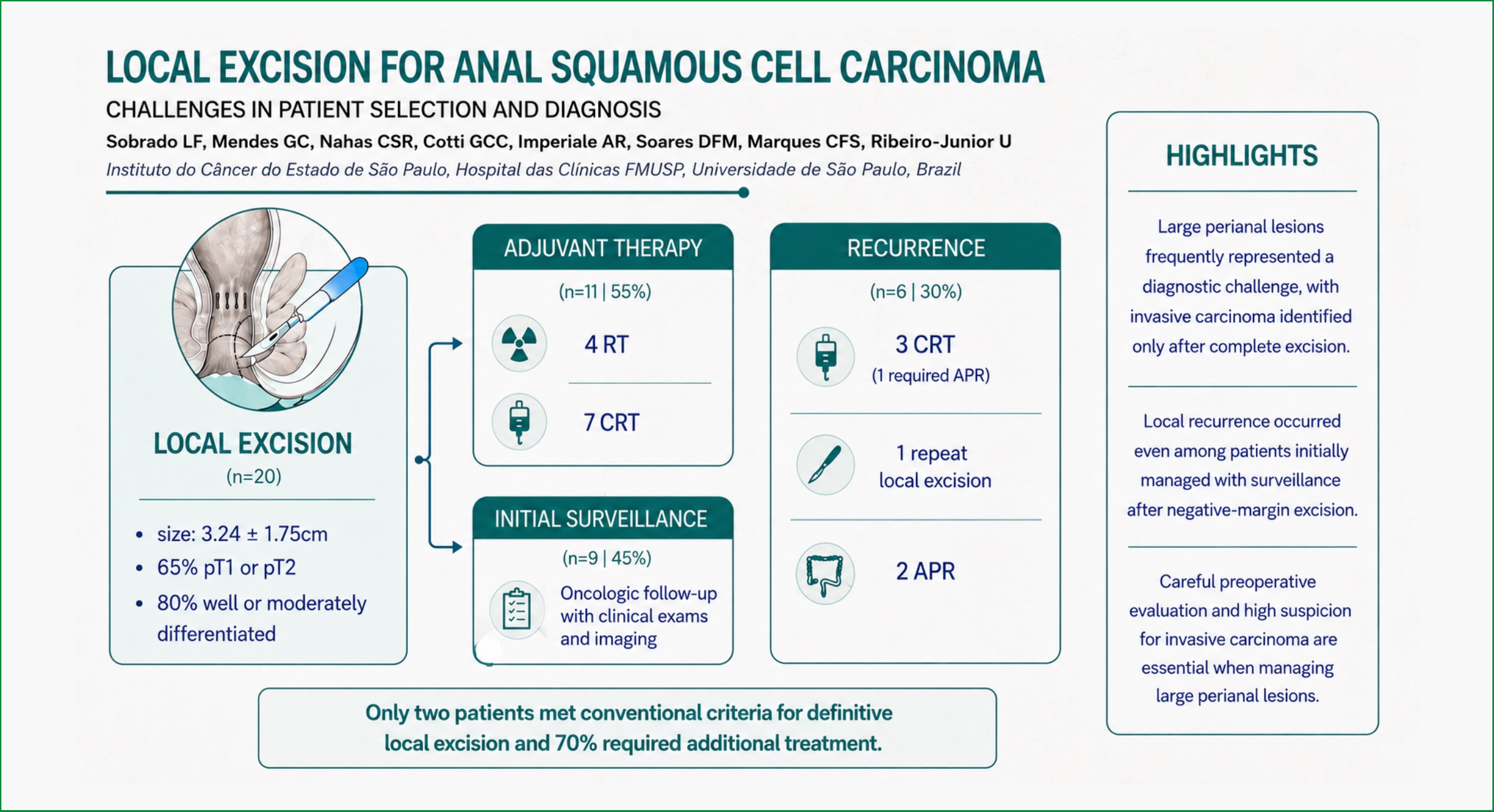
Local excision (LE) has a limited role in the management of anal squamous cell carcinoma (SCC), typically restricted to carefully selected early-stage tumors. However, in clinical practice, LE is often performed in broader contexts, particularly in patients with large perianal lesions without prior confirmation of invasive carcinoma, in whom diagnostic uncertainty may influence treatment decisions.
To evaluate oncologic outcomes after LE for anal SCC, with particular emphasis on the role of lesion characteristics, diagnostic uncertainty, and treatment patterns in real-world practice.
This was a retrospective study of patients with anal SCC who underwent LE as primary treatment and were followed at a tertiary cancer center between 2010 and 2024. Clinical, pathological, and treatment data were collected, including tumor characteristics, margin status, use of adjuvant therapy, and oncologic outcomes.
A total of 20 patients with invasive anal SCC were included. The mean tumor size was 3.24 cm (standard deviation ±1.75 cm), with lesions up to 6.9 cm. No patients received chemoradiotherapy before LE. Only two patients met conventional criteria for LE (<2 cm, well or moderately differentiated tumors). Overall, 14 patients (70%) required additional treatment following LE, including chemoradiotherapy or abdominoperineal resection. Among patients initially managed with surveillance, 44% developed local recurrence, even in cases with negative margins.
Outcomes following LE for anal SCC are strongly influenced by patient selection and clinical context. In this real-world cohort, most patients outside established criteria required additional treatment, underscoring the limitations of LE in large perianal lesions. These findings highlight the importance of careful preoperative evaluation and maintaining a high index of suspicion for invasive carcinoma to guide appropriate initial management.
Early-onset colorectal cancer (EOCRC), defined as diagnosis before 50 years of age, has increased in incidence globally, but its clinicopathologic profile and prognostic significance remain incompletely characterized, particularly in Southeast Asian populations with limited access to organized screening.
To compare clinicopathologic characteristics and survival outcomes between EOCRC and late-onset colorectal cancer (LOCRC) following curative resection, and to identify independent prognostic determinants in a regional Vietnamese surgical cohort.
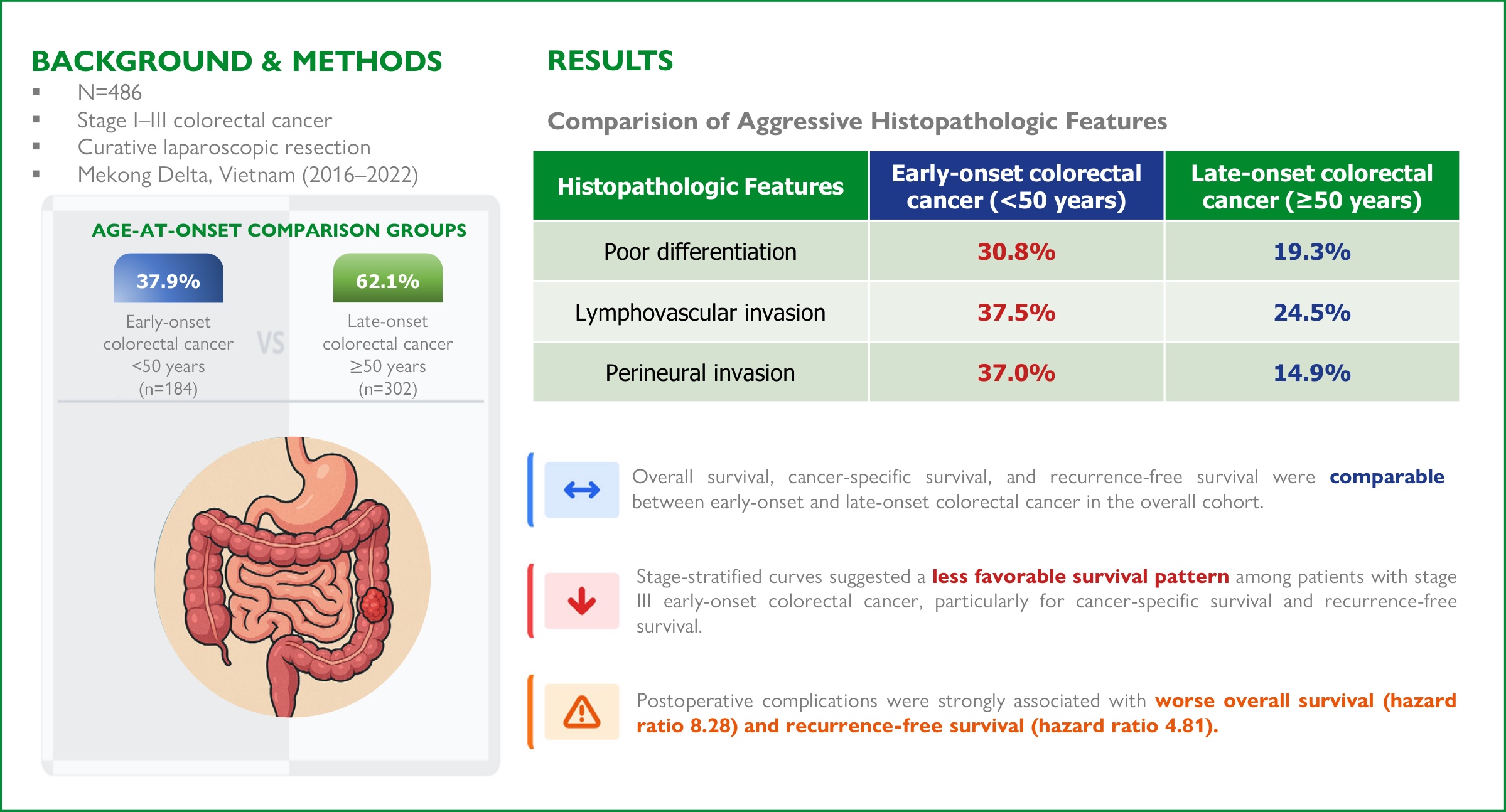
This retrospective cohort study included 486 patients with stage I-III colorectal adenocarcinoma who underwent curative laparoscopic resection at a tertiary center in the Mekong Delta between 2016 and 2022.
EOCRC (184 patients, 37.9%) was associated with higher rates of poor differentiation, mucinous or signet-ring cell histology, lymphovascular invasion, and perineural invasion compared with LOCRC (all p<0.05). Overall survival (OS), cancer-specific survival (CSS), and recurrence-free survival (RFS) did not differ significantly between groups. In the overall cohort (log-rank p=0.72, 0.15, and 0.96, respectively). On multivariable analysis, postoperative complications were the dominant independent prognostic factor for OS (hazard ratio [HZ] 8.28; 95% confidence interval [CI] 4.87-14.09) and RFS (HZ 4.81; 95%CI 3.03-7.65), whereas age at onset was not independently associated with either outcome. Stage-stratified curves suggested a less favorable survival pattern among patients with stage III EOCRC, particularly for CSS and RFS.
Despite a more aggressive histopathologic profile, EOCRC showed comparable survival to LOCRC in the overall cohort. Stage-stratified curves suggested a less favorable survival pattern among patients with stage III EOCRC, while postoperative complications represented the dominant modifiable determinant of outcome.
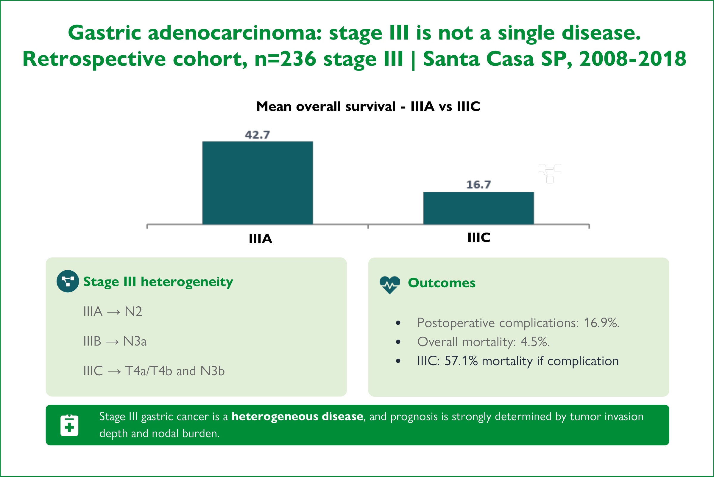
Gastric adenocarcinoma remains one of the leading causes of cancer mortality worldwide, according to the International Agency for Research on Cancer. Stage III, as defined by the 8th edition of the American Joint Committee on Cancer Tumor, Node, and Metastasis (TNM) system, comprises a biologically heterogeneous group, resulting in clinicopathological variations that directly impact prognosis and therapeutic decisions.
To evaluate the clinicopathological features, surgical morbidity and mortality, and overall survival of patients with gastric adenocarcinoma treated at a tertiary center, and to analyze prognostic differences among stage III subgroups.
This retrospective cohort study included patients treated between 2008 and 2018. Demographic variables, tumor characteristics, TNM staging (8th edition), type of surgical procedure, complications according to the Clavien-Dindo classification, and 30-day mortality were analyzed. Overall survival was estimated using the Kaplan-Meier method and compared with the log-rank test, with a significance level of 5%.
This retrospective cohort study included patients treated between 2008 and 2018. We analyzed demographic variables, tumor characteristics, TNM staging (8th edition), type of surgical procedure, complications according to the Clavien-Dindo classification, and 30-day mortality. Overall survival was estimated using the Kaplan-Meier method and compared with the log-rank test, with a significance level of 5%.
Stage III gastric adenocarcinoma demonstrates marked clinicopathological heterogeneity, which directly impacts morbidity, mortality, and survival. Lymph node involvement is a relevant prognostic determinant.




Developed by Surya MKT