

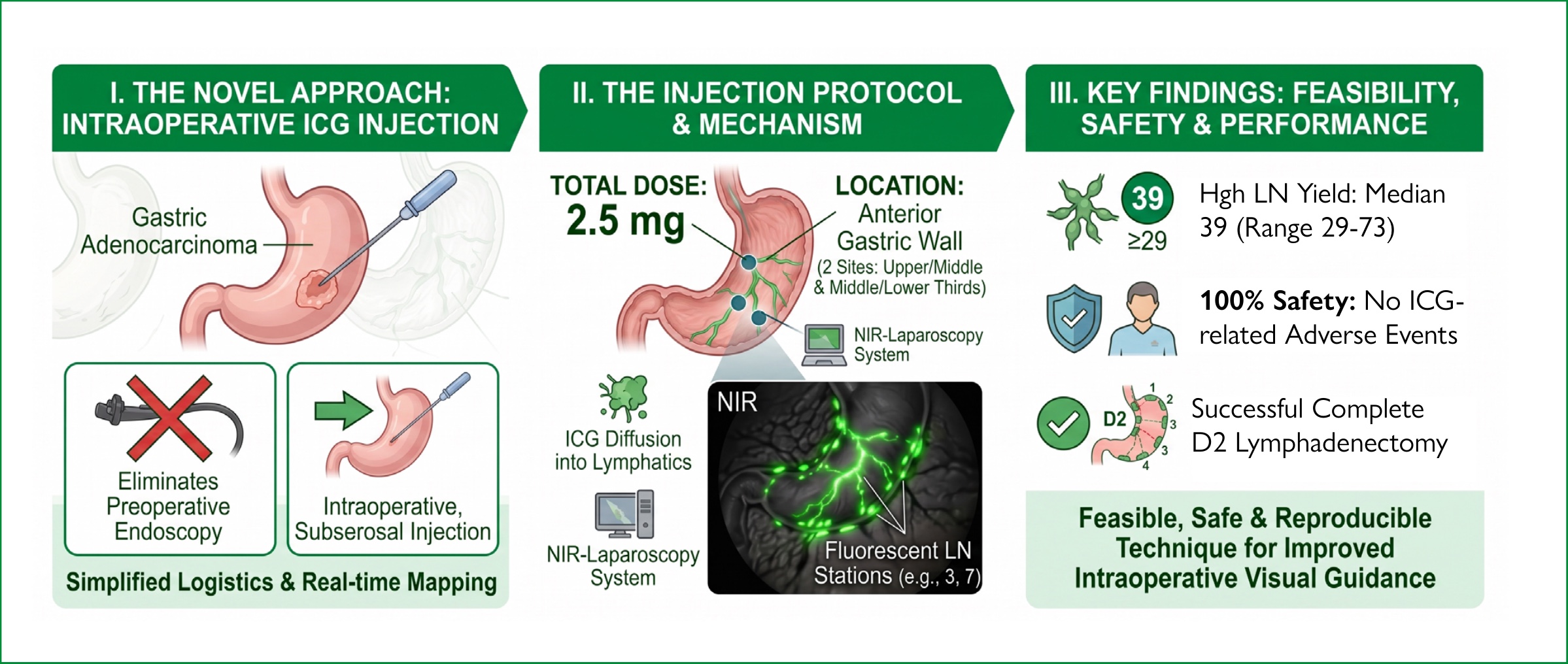
Fluorescence-guided lymphadenectomy using indocyanine green (ICG) improves intraoperative visualization of gastric lymphatic drainage during gastrectomy. Most reported techniques rely on preoperative endoscopic injection, and standardized intraoperative protocols are limited.
To evaluate the feasibility, safety, and lymph node retrieval of intraoperative subserosal ICG injection for fluorescence-guided D2 lymphadenectomy during laparoscopic gastrectomy.
A prospective descriptive pilot study was conducted at two institutions. Consecutive patients with gastric adenocarcinoma undergoing laparoscopic total or subtotal gastrectomy with curative intent and D2 lymphadenectomy were included. ICG was injected intraoperatively into the anterior gastric wall (two injections of 1.25 mg) after diagnostic exploration. Outcomes included feasibility, safety, fluorescence distribution across D2 stations, and total lymph node retrieval.
Twenty patients were included. The protocol was successfully implemented in all cases without technical difficulties or ICG-related adverse events. Complete D2 lymphadenectomy was achieved in 100% of patients. The median number of retrieved lymph nodes was 39 (range 29–73), with =30 nodes in 95% of cases. Fluorescent nodes were consistently identified in stations 3, 4, 6, and 7, with variable distribution in stations 5, 9, 8a, 11, 1, and 2, and absent in station 12a.
Intraoperative subserosal ICG injection is a feasible, safe, and reproducible technique that enables consistent visualization of major gastric lymphatic basins without requiring preoperative endoscopic injection.
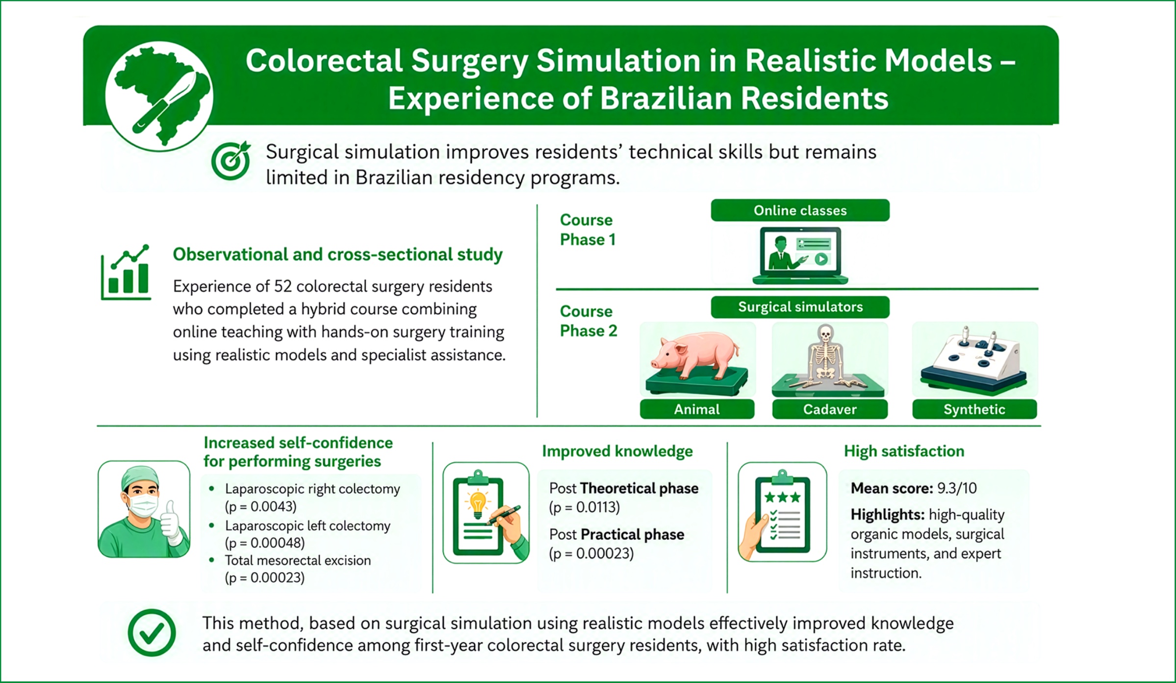
Practical training is essential in surgical education, and simulation is an effective strategy for developing technical skills in a safe environment. This study describes the experience of the Advanced Practical Course in Colorectal Surgery, which enrolled 52 colorectal surgery residents from different regions of Brazil. The course provided supervised training in colorectal procedures using realistic models, including synthetic simulators, animal models, and fresh cadavers.
To evaluate the applicability of a theoretical-practical training method for first-year colorectal surgery residents in Brazil, as well as theoretical knowledge acquisition, self-confidence in performing procedures, and participant satisfaction.
This study describes the experience of the Advanced Practical Course in Colorectal Surgery, which enrolled 52 colorectal surgery residents from different regions of Brazil. The course provided supervised training in colorectal procedures using realistic models, including synthetic simulators, animal models, and fresh cadavers.
The course was divided into two phases: (1) theoretical lectures and intraoperative videos delivered through a live online platform; and (2) two days of surgical simulation using cadaveric, porcine, and synthetic models under the supervision of experienced colorectal surgeons. Objective theoretical assessments were performed before and after each phase, along with subjective evaluations at the beginning and end of the course. Statistical analysis was conducted using R Commander® software. A significant improvement in self-confidence was observed for laparoscopic right colectomy (p=0.0043), laparoscopic left colectomy (p=0.00048), and total mesorectal excision (p=0.00023). Technical knowledge increased after both the theoretical (p=0.0113) and practical (p=0.00023) phases. Overall satisfaction was high, with a mean score of 9.3 out of 10.
The theoretical-practical simulation-based method proved to be applicable and effective, leading to improved knowledge acquisition, increased procedural confidence, and high satisfaction among first-year colorectal surgery residents.
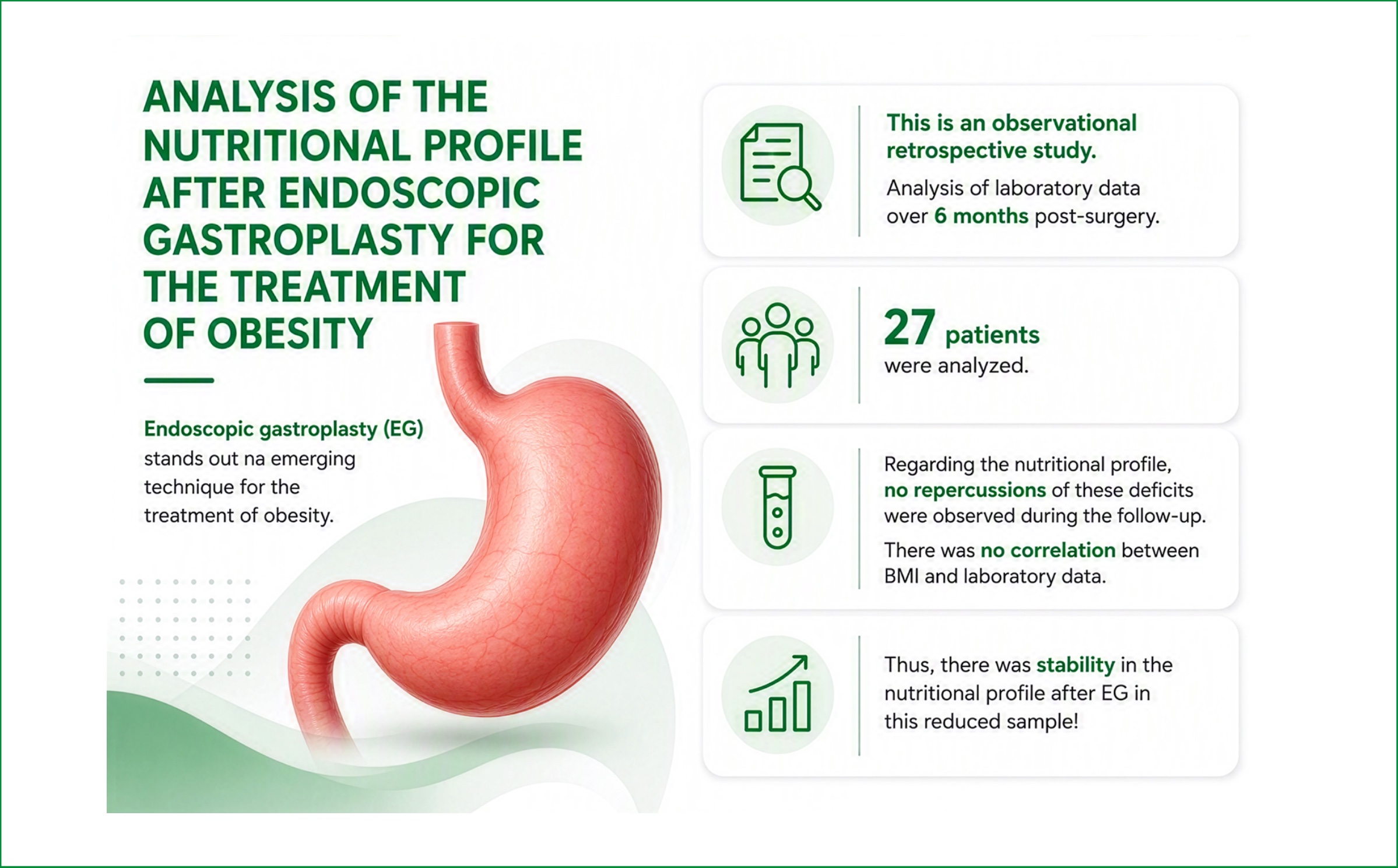
Endoscopic gastroplasty (EG) has emerged as a promising technique for the treatment of obesity. However, data on the nutritional profile of patients after this surgical procedure are scarce in the literature.
To analyze the nutritional profile of patients who underwent EG for the treatment of obesity during the postoperative period.
This retrospective observational study investigated medical record data from a private healthcare service in Salvador, Bahia, where EG was performed EG. Epidemiological and laboratory data (total protein, albumin, calcium, zinc, vitamin B12, 25-hydroxyvitamin D, hemoglobin, glucose, glycated hemoglobin, ferritin, and iron) were collected before and after surgery over a 6-month period. A p-value>0.05 was considered statistically significant.
Twenty-seven patients were analyzed, with a mean age of 43.8 (±13.3) years, of whom 88.9% were female. The patients’ initial body mass index (BMI) was 35.31±8.7 kg/m2. Total weight loss was 10±5.7, and excess weight loss was 41.1±28.1. Preoperatively, iron deficiency was observed in 1 patient, vitamin B12 deficiency in 2 patients, 25-hydroxyvitamin D deficiency in 2 patients, zinc deficiency in 1 patient, calcium deficiency in 2 patients, and hyperglycemia in 4 patients. No consequences of these deficiencies were observed during follow-up. There was no correlation between BMI and laboratory data (p>0.05).
The nutritional profile remained stable before and after EG, with sustained weight loss.
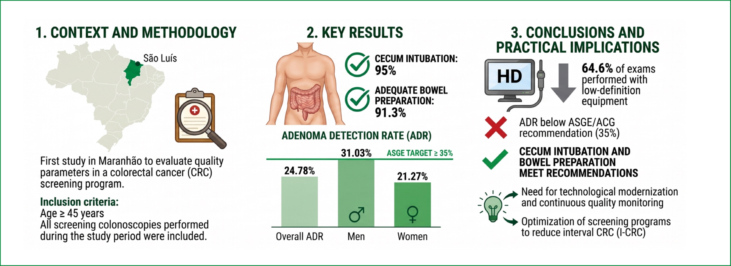
Colorectal cancer is the third most frequently diagnosed cancer worldwide. Colonoscopy is fundamental in reducing its incidence and mortality, provided it follows certain quality parameters. Examples of these quality parameters are the cecal intubation rate, adenoma detection rate (ADT), and the adequate percentage of patients with satisfactory bowel preparation.
To measure the quality parameters of a colonoscopy service in Northeast Brazil.
A cross-sectional, retrospective study, with data collected from electronic medical records, including screening colonoscopies between 2022 and 2024.
565 colonoscopies were included in the study. The overall ADR was 24.78, 21.27% in women and 32.03% in men. The cecal intubation rate was approximately 95%, and most patients (91.3%) had excellent bowel preparation. The polyp detection rate was 39.47%.
The cecal intubation rate and the proportion of patients with adequate bowel preparation were satisfactory. However, the polyp detection rate was lower than recommended in the literature. Thus, evaluating these parameters is a way to monitor the quality of colorectal cancer prevention, improving the strategies employed, even in the face of the limitations encountered.
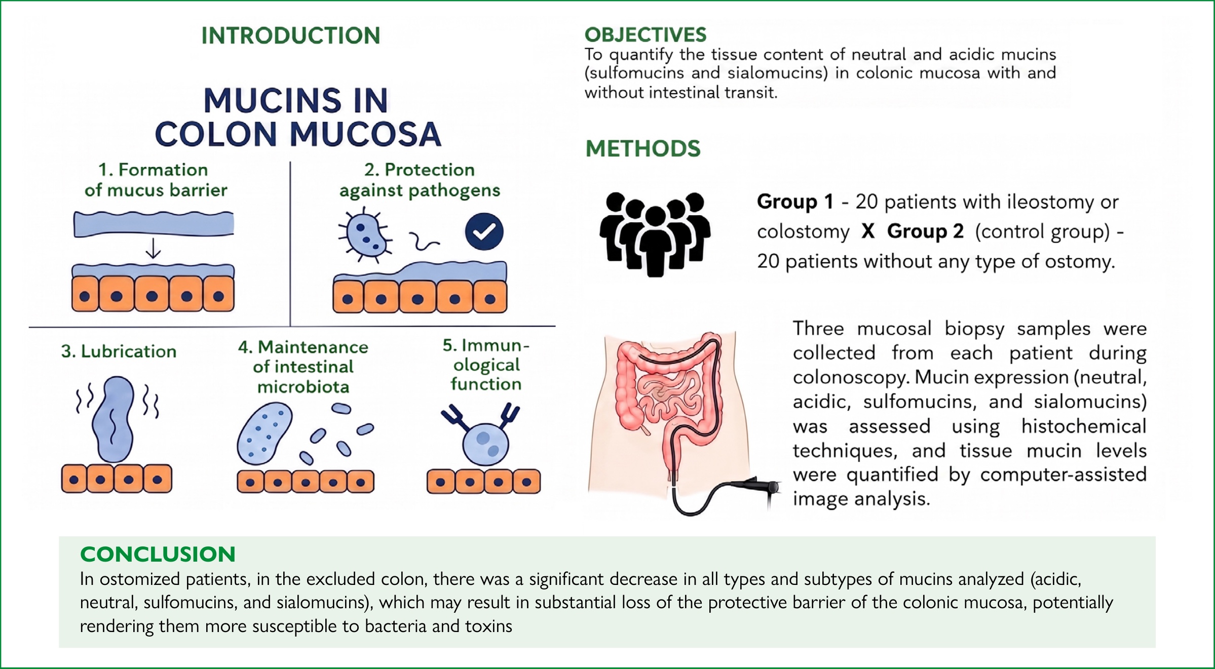
Alterations in mucin expression in the intestinal mucosa are among the main histological findings in inflammatory colonic diseases. No previous study has quantified the tissue expression of neutral and acidic mucins, including sulfomucins and sialomucins, in patients with diversion colitis.
To quantify the tissue content of neutral and acidic mucins (sulfomucins and sialomucins) in colonic mucosa with and without intestinal transit.
Forty patients undergoing elective colonoscopy at the Hospital de Clínicas – Unicamp were included. Group 1 comprised 20 patients with an ileostomy or colostomy for more than two months. Group 2 (control) included 20 patients without any type of ostomy. Patients with inflammatory bowel disease or previous pelvic radiotherapy were excluded. Three mucosal biopsies were obtained from each patient during colonoscopy. Neutral and acidic mucins were assessed using Periodic Acid-Schiff and Alcian Blue staining, respectively. Acidic mucin subtypes (sulfomucins and sialomucins) were identified using Alcian Blue-high iron diamine. Tissue content was quantified by computer-assisted image analysis. Statistical analysis compared mucin levels between segments with and without fecal transit, with significance set at p<0.05.
Colonic segments without fecal transit showed a significant reduction in the tissue content of all mucin subtypes.
All mucin subtypes (neutral, acidic, sulfomucins, and sialomucins) were significantly reduced in colonic segments deprived of fecal stream.
Colorectal cancer is a global health problem with high incidence and mortality rates, frequently diagnosed at metastatic stages. Mutations in the KRAS gene play a key role in colorectal cancer progression, significantly influencing treatment resistance and prognosis.
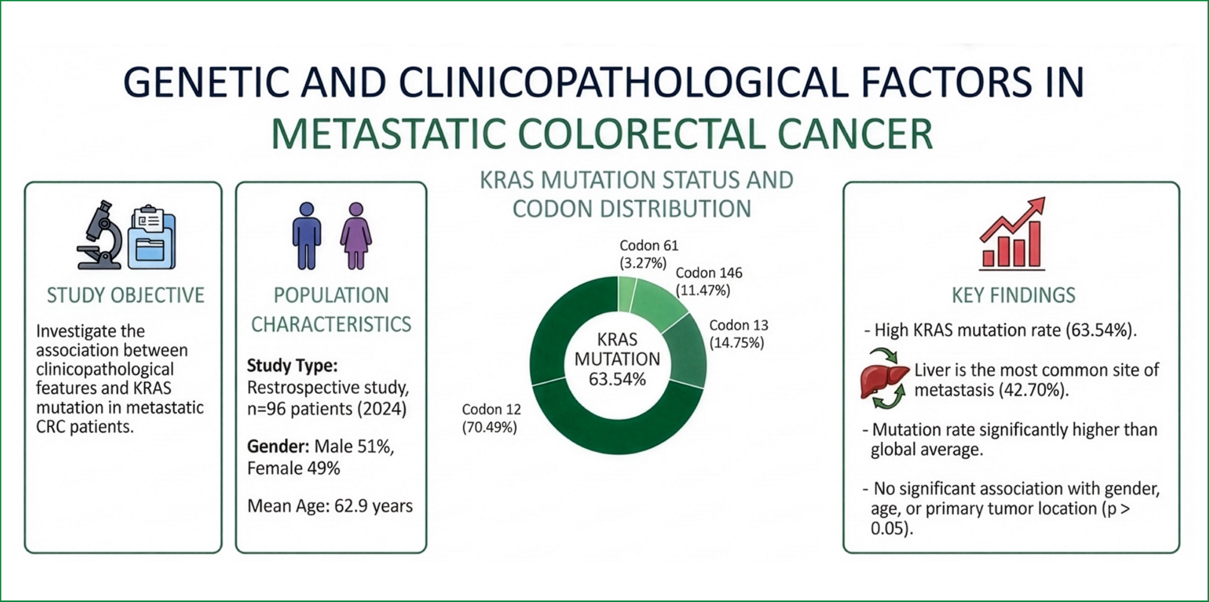
This study investigated the association between clinicopathological characteristics and KRAS mutations in patients with metastatic colorectal cancer.
This retrospective study evaluated medical records of patients with metastatic colorectal carcinoma who underwent KRAS gene mutation testing. A total of 96 patients treated in 2024 were included in the analysis.
KRAS mutations were identified in 63.54% of patients, predominantly in codon 12 (70.49%) and codon 13 (14.75%). No significant associations were found between KRAS mutation status and factors such as sex, age, or tumor location. Nevertheless, KRAS mutations were more frequently observed in patients with left-sided colon tumors, contrasting with the findings described in the literature. KRAS mutations were associated with poorer prognosis, higher rates of metastases (particularly to the liver), and higher rates of resistance to conventional treatments, such as chemotherapy.
This study highlights the importance of KRAS mutation testing to support personalized, more effective treatment strategies to improve the prognosis of patients with metastatic colorectal cancer.
Bronchopulmonary aspiration occurs when food or fluids enter the lower airways, potentially causing hypoxia, aspiration pneumonia, and even death. Prevention requires early identification of risks and action by a multidisciplinary team. Continuous team education, using well established protocols, is essential to reinforce preventive measures and improve healthcare.
Based on the well-established protocols, evaluate the impact of training on professional learning and the incidence of bronchopulmonary aspiration.
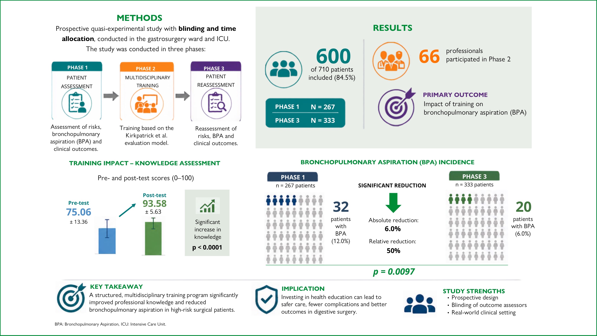
A prospective, blinded, time-allocated, quasi-experimental study conducted in the gastrointestinal surgery ward and intensive care unit. The study was divided into three phases: patient assessment, multidisciplinary training based on the well-established protocols, and patient reassessment regarding risks, bronchopulmonary aspiration, and outcomes.
The study included 600 of the 710 patients evaluated, distributed in two phases: Phase 1 (n=267) and Phase 3 (n=333). Sixty-six professionals participated in Phase 2. The impact of training on bronchopulmonary aspiration was assessed through pre- and post-training assessments. The score increased significantly (pre-test: 75.06±13.36; post-test: 93.58±5.63; p<0.0001). In Phase 1, 32 patients presented with bronchopulmonary aspiration; in Phase 3, this number was 20, a significant difference (p=0.0097).
Training proved effective in preventing bronchopulmonary aspiration and improving clinical outcomes, with a positive impact at all levels of the protocol employed. The results reinforce continuing education as a strategy for patient safety and risk management, suggesting its application in other healthcare settings.
Roux-en-Y gastric bypass promotes weight loss in patients with severe obesity and improves type 2 diabetes mellitus, which, in some cases, can lead to remission. The action of enteric hormones is relevant for the initial outcomes and late disease evolution.
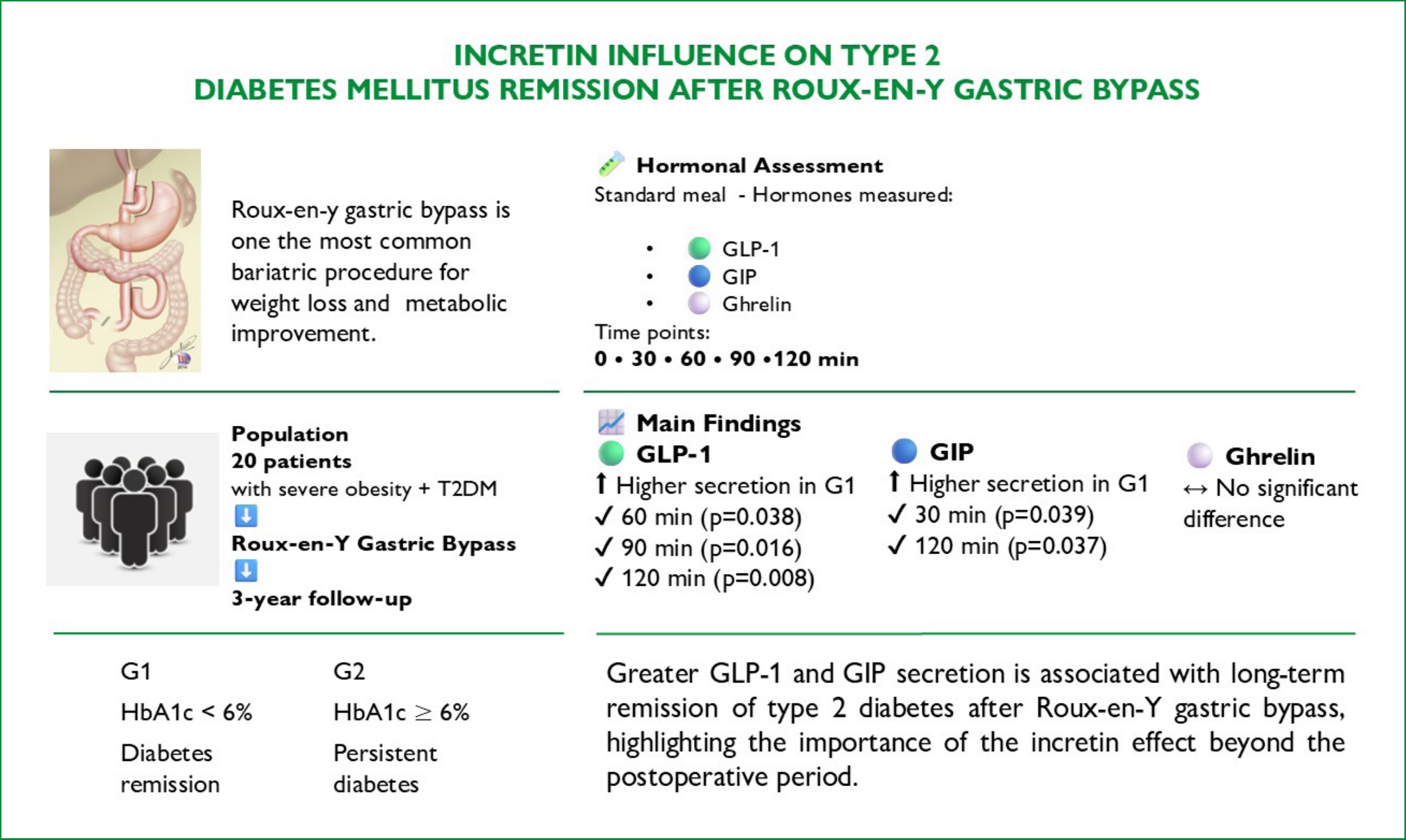
To evaluate the influence of enteric hormone secretion on the evolution of type 2 diabetes mellitus three years after Roux-en-Y gastric bypass.
A total of 20 patients were evaluated, divided into 2 groups: group 1, glycated hemoglobin<6%, and group 2, glycated hemoglobin≥6%. Hormone secretion (ghrelin, GLP-1, and GIP) was analyzed at pre-specified time points following a standard meal.
Group 1 had higher mean secretion at 30 and 120 minutes ([992.60±806.78 vs. 407.97±186.80, p=0.039, p<0.05] and [418.72±219.11 vs. 240.14±121.11, p=0.037, p<0.05], respectively) than group 2. Group 1 presented higher GLP values at 60 [724 (195–1,483) vs. 214 (67–424), p=0.038, p<0.05], 90 [473 (200–943) vs. 180 (120–318), p=0.016, p<0.05] and 120 minutes [565 (278–770) vs. 163 (81–339), p=0.008, p<0.05] compared to group 2. There was no difference between the groups regarding ghrelin.
Higher secretion of GIP and GLP-1 was observed in patients with type 2 diabetes mellitus remission. The incretin factor is important in late disease evolution even in a group of patients with severe diabetes.
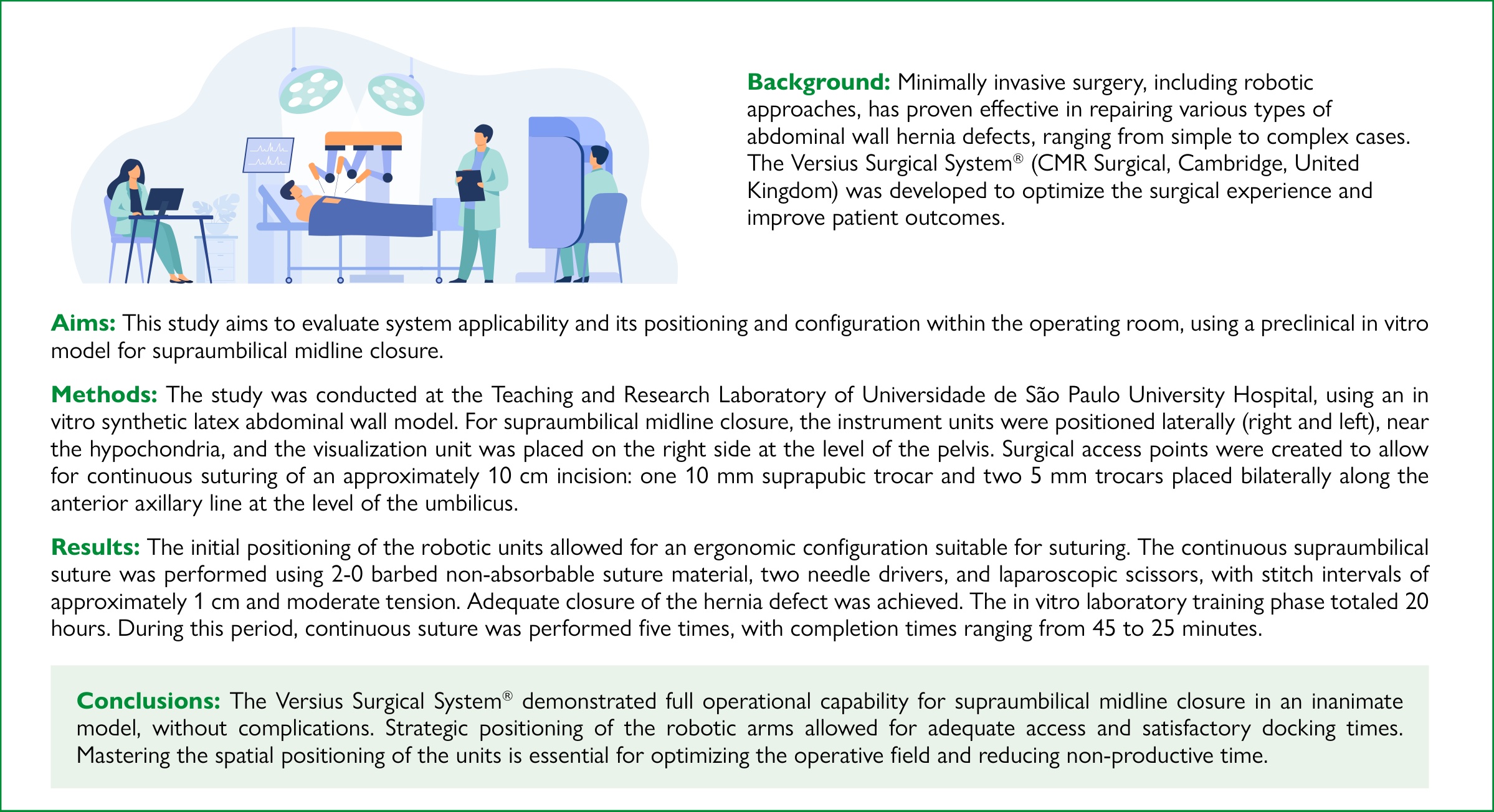
Minimally invasive surgery, including robotic approaches, has proven effective in repairing various types of abdominal wall hernia defects, ranging from simple to complex cases. The Versius Surgical System® (CMR Surgical, Cambridge, United Kingdom) was developed to optimize the surgical experience and improve patient outcomes.
This study aims to evaluate system applicability and its positioning and configuration within the operating room, using a preclinical in vitro model for supraumbilical midline closure.
The study was conducted at the Teaching and Research Laboratory of Universidade de São Paulo University Hospital, using an in vitro synthetic latex abdominal wall model. For supraumbilical midline closure, the instrument units were positioned laterally (right and left), near the hypochondria, and the visualization unit was placed on the right side at the level of the pelvis. Surgical access points were created to allow for continuous suturing of an approximately 10 cm incision: one 10 mm suprapubic trocar and two 5 mm trocars placed bilaterally along the anterior axillary line at the level of the umbilicus.
The initial positioning of the robotic units allowed for an ergonomic configuration suitable for suturing. The continuous supraumbilical suture was performed using 2-0 barbed non-absorbable suture material, two needle drivers, and laparoscopic scissors, with stitch intervals of approximately 1 cm and moderate tension. Adequate closure of the hernia defect was achieved. The in vitro laboratory training phase totaled 20 hours. During this period, continuous suture was performed five times, with completion times ranging from 45 to 25 minutes.
The Versius Surgical System® demonstrated full operational capability for supraumbilical midline closure in an inanimate model, without complications. Strategic positioning of the robotic arms allowed for adequate access and satisfactory docking times. Mastering the spatial positioning of the units is essential for optimizing the operative field and reducing non-productive time.
Clinical features and outcomes of patients admitted to the intensive care unit due to acute abdomen are important to be investigated.
To evaluate the outcomes of critically ill subjects with acute abdomen according to etiology, comorbidity and severity.
Outcomes of 1,523 patients (878 women, mean age 66±18 years) consecutively admitted to a specialized gastrointestinal intensive care unit with different causes of acute abdomen from January 2012 to December 2019, were retrospectively evaluated according to etiology, comorbidity and severity.
The most common causes of acute abdomen were obstructive and inflammatory, particularly large bowel obstruction (27%), small bowel obstruction (18%) and acute pancreatitis (17%). Overall mortality was 13%. Surgery was required in 34% of patients. Median length of stay in the hospital was 9 [1-101] days. On univariate analysis mortality was significantly associated with age, APACHE II, Charlson comorbidity index, requirement for surgery and malignancy (p<0.0001), but only APACHE II, Charlson comorbidity index and surgical interventional remained significant on multivariate analysis.
Critically ill patients admitted to the intensive care unit with acute abdomen constitute a heterogeneous group of subjects with different prognosis. Mortality is more related to the severity of the disease, comorbidity and need for surgery than to the etiology of the acute abdomen.




Developed by Surya MKT