

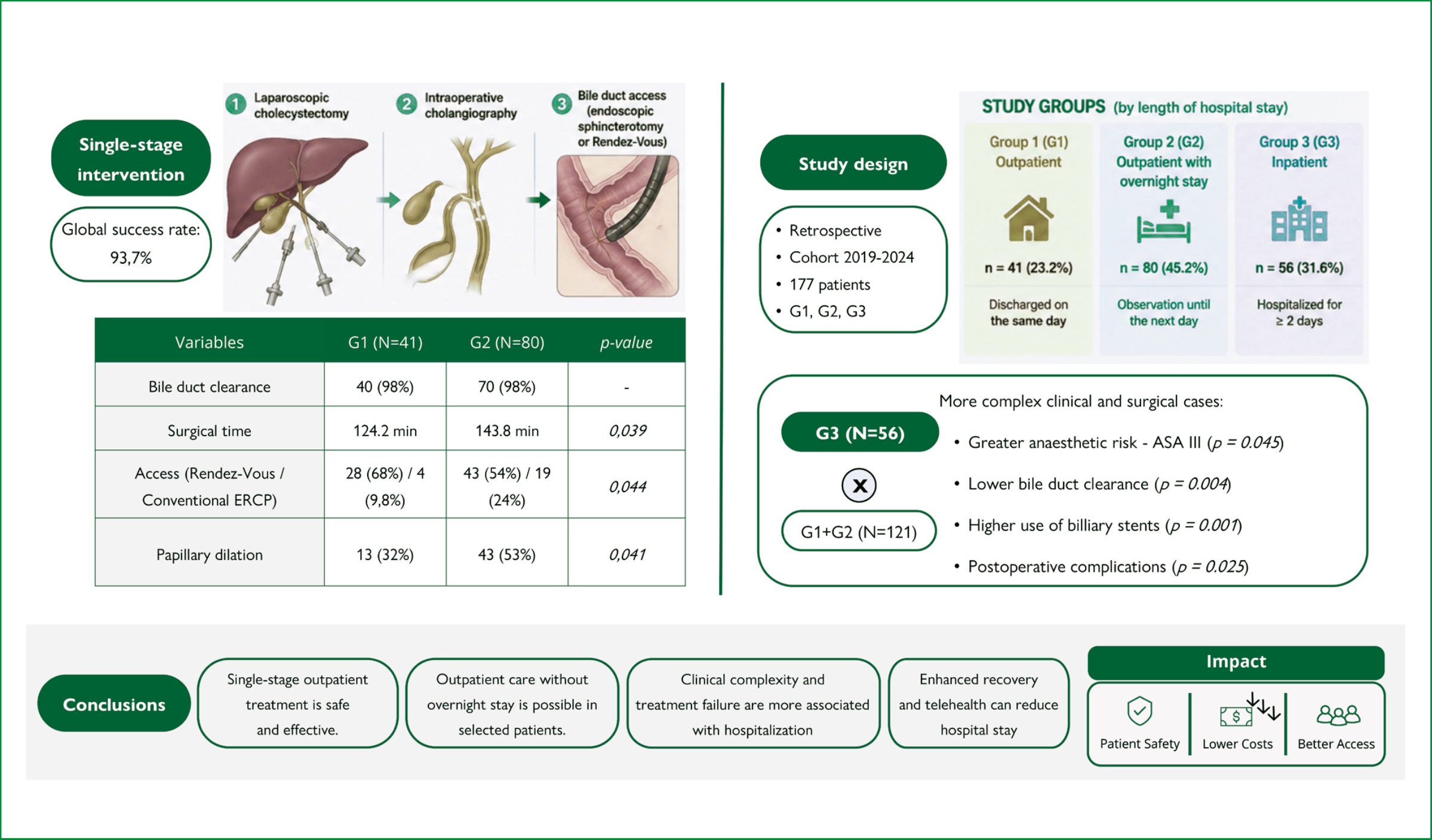
Single-stage outpatient treatment of cholecystocholedocholithiasis is feasible, highlighting the importance of appropriate patient selection, professional training, and healthcare service organization to support this approach.
To identify clinical and procedural factors associated with outpatient management and hospital stay following single-stage laparoendoscopic treatment of cholecystocholedocholithiasis.
A retrospective cohort study was conducted at hospitals affiliated with the Ribeirão Preto School of Medicine, Universidade de São Paulo (FMRP-USP), between 2019 and 2024. Patients were stratified into three groups according to care setting: outpatient (G1); outpatient with overnight stay (G2); and inpatient (G3). Clinical, surgical, and outcome data were analyzed using logistic regression models, χ2 tests, and Fisher’s exact tests (p=0.05).
Among 177 patients included, 41 were allocated to G1, 80 to G2, and 56 to G3. Compared with G2, G1 patients had shorter operative time (124.2 vs. 143.8 min; p=0.038), more frequent use of the Rendez-Vous technique (p=0.044), and less frequent use of papillary dilation (p=0.041). Patients in G3 had a higher prevalence of ASA III physical status (p=0.045), higher rates of postoperative complications (p=0.025) and biliary stent use (p=0.001), and lower bile duct clearance rates (p=0.004).
Single-stage outpatient laparoendoscopic treatment of cholecystocholedocholithiasis is safe and effective. Clinical severity, treatment complexity, and failure of bile duct clearance were more strongly associated with inpatient hospitalization, whereas overnight stay among outpatients was not associated with improved postoperative outcomes. The implementation of enhanced recovery protocols and telemedicine-based postoperative assessment strategies may further reduce hospital stay.
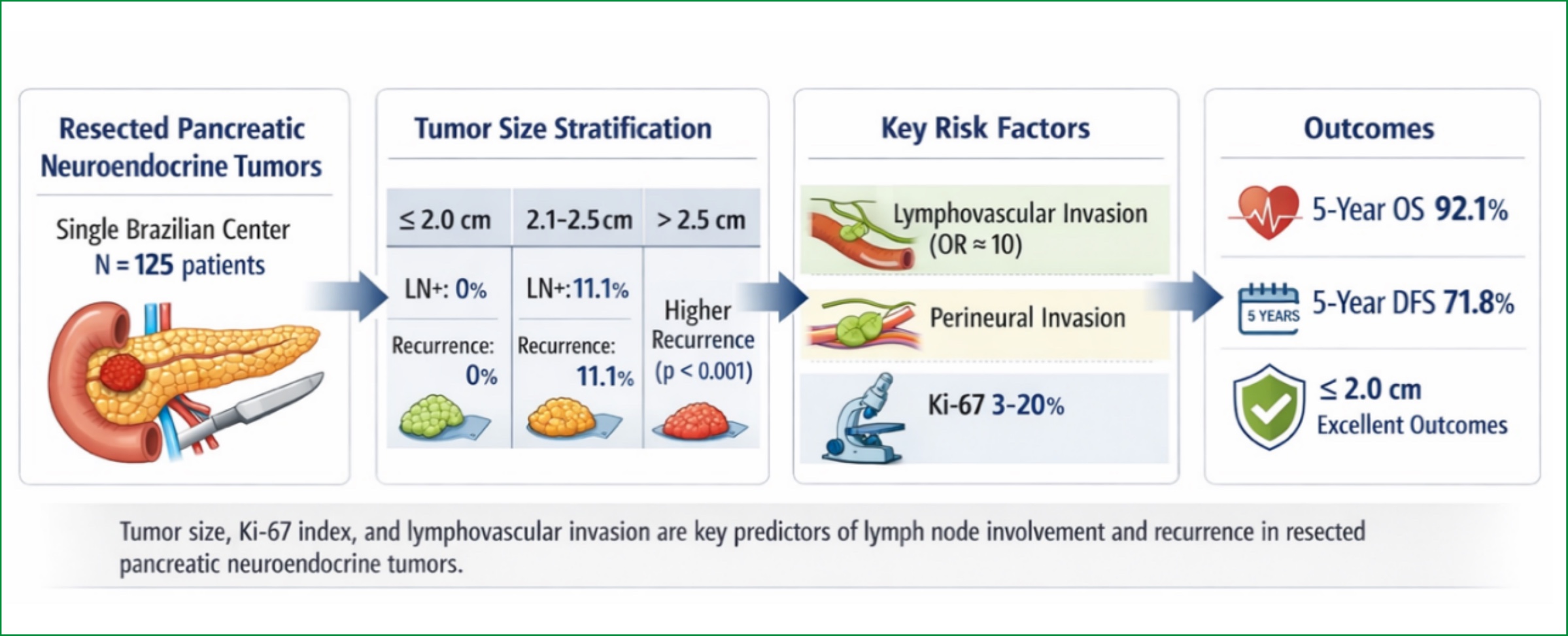
Pancreatic neuroendocrine tumors (pNETS) constitute a heterogeneous and rare disease worldwide. Diagnosis rates have been increasing, and the evaluation of prognostic factors has become even more important in the treatment decision-making process.
To present results and prognostic factors in patients undergoing surgical treatment for pNETS at a single Brazilian center.
The most frequently performed surgical procedure was PCC+S 60.0%. The median hospital stay was 8 (5-13) days, and postoperative mortality occurred in 1.7%. Overall survival in 3 and 5 years was 93.8 and 92.1%, respectively. Disease-free survival at 3 and 5 years was 87.1 and 71.8%, respectively. Patients with tumors smaller than 2.0 cm did not present with lymph node disease or recurrence, and patients with tumors measuring 2.1-2.5cm had lymph node disease in 11.1% and recurrence in 11.1%. In univariate and multivariate analysis, the presence of lymphatic and perineural invasion and Ki67 (3-20) were strongly correlated with positive lymph node disease and recurrence, respectively.
The presence of lymphatic and perineural invasion, Ki67 (3 to 20), and tumors larger than 2.5 cm correlated with positive lymph node disease and recurrence.
Obesity is a multifactorial disease with a high prevalence that leads to several comorbidities, posing significant challenges for healthcare systems. Bariatric and metabolic surgery (BMS) has been established as the most effective treatment for patients with obesity; however, in Brazil, limited access remains a critical barrier.
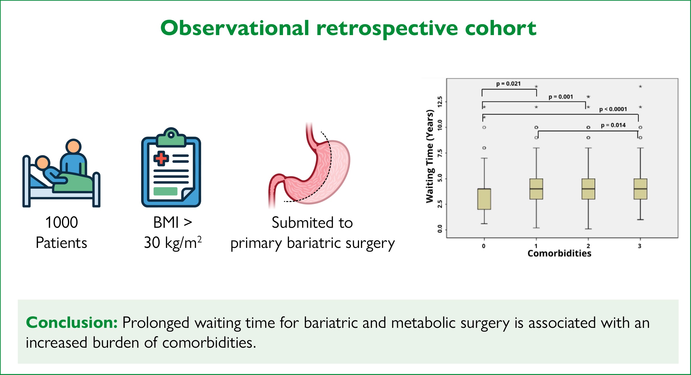
This study aimed to evaluate the clinical, demographic, and metabolic characteristics of patients with obesity undergoing BMS in the Brazilian Unified Health System and to analyze the relationship between waiting time and comorbidities.
A retrospective cohort study was conducted involving 1,000 patients with obesity who underwent treatment between July 2022 and June 2024. Clinical, anthropometric, and laboratory variables were analyzed using regression analysis and statistical tests to assess the association between waiting time and comorbidities.
A significant correlation was found between prolonged waiting time and an increased number of comorbidities (R²=0.686; p<0.001). Furthermore, the number of comorbidities explained 60% of the variability in waiting time (R²=0.600; p<0.001), with a mean increase of 1.92 years for each additional comorbidity (95%CI 1.82–2.02). Patients on the waiting list for more than 10 years had higher rates of hypertension, type 2 diabetes, and dyslipidemia. Waiting time also had an impact on some metabolic syndrome parameters, including glycated hemoglobin (Hb1Ac) (r=+680, p=0.031), low-density lipoprotein (LDL) (r=+640, p=0.044), and total cholesterol (r=+830, p=0.008).
Prolonged waiting time for bariatric and metabolic surgery is associated with an increased burden of metabolic comorbidities and their consequences.
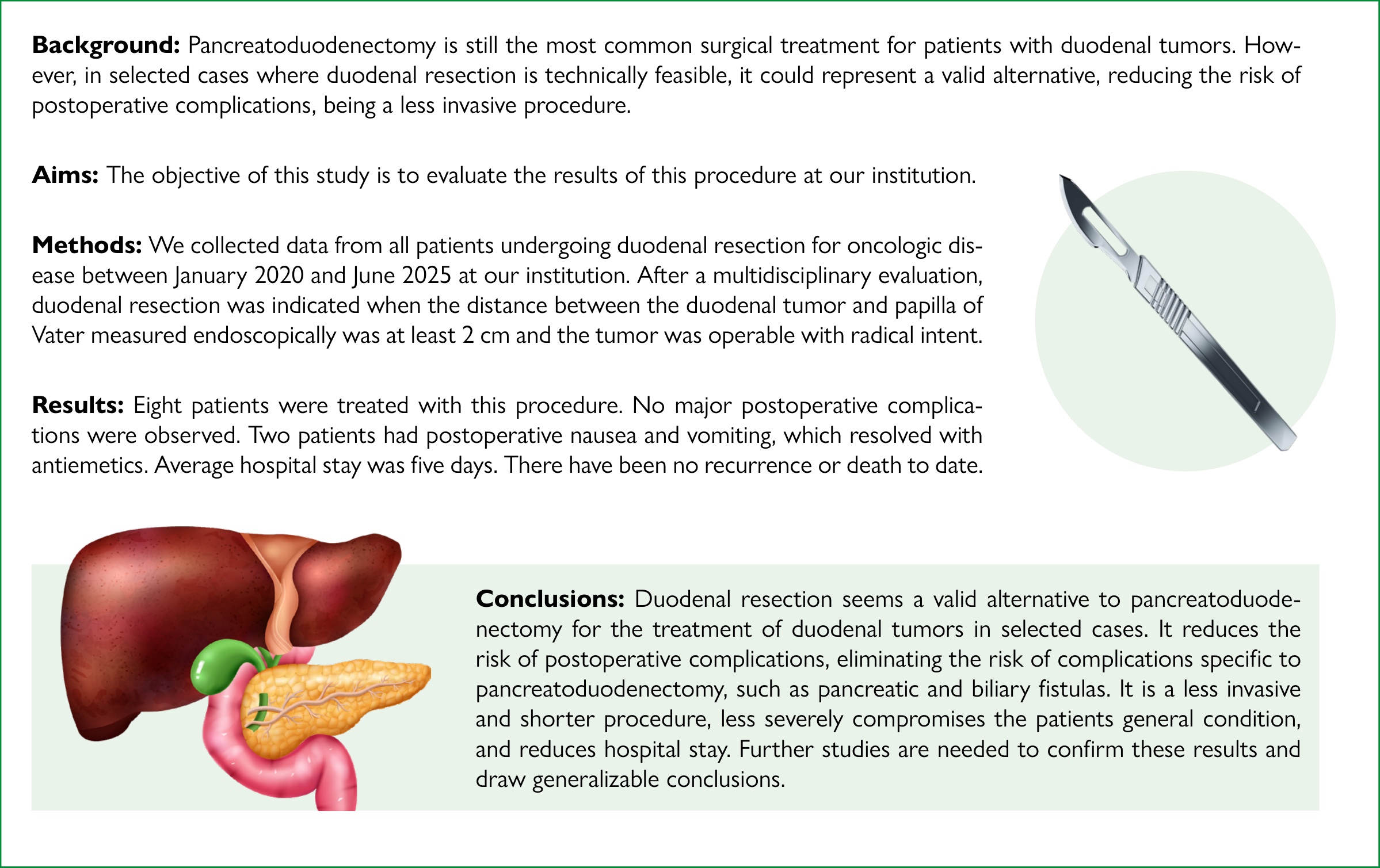
Pancreatoduodenectomy is still the most common surgical treatment for patients with duodenal tumors. However, in selected cases where duodenal resection is technically feasible, it could represent a valid alternative, reducing the risk of postoperative complications, being a less invasive procedure.
The objective of this study was to evaluate the results of this procedure at our institution.
We collected data from all patients undergoing duodenal resection for oncologic disease between January 2020 and June 2025 at our institution. After a multidisciplinary evaluation, duodenal resection was indicated when the distance between the duodenal tumor and papilla of Vater measured endoscopically was at least 2 cm, and the tumor was operable with radical intent.
Eight patients were treated with this procedure. No major postoperative complications were observed. Two patients had postoperative nausea and vomiting, which resolved with antiemetics. Average hospital stay was five days. There has been no recurrence or death to date.
Duodenal resection seems a valid alternative to pancreatoduodenectomy for the treatment of duodenal tumors in selected cases. It reduces the risk of postoperative complications, eliminating the risk of complications specific to pancreatoduodenectomy, such as pancreatic and biliary fistulas. It is a less invasive and shorter procedure, less severely compromises the patients general condition, and reduces hospital stay. Further studies are needed to confirm these results and draw generalizable conclusions.
Removal of malignant tumors with free margins is pivotal in oncological surgery.
To verify the correspondence between the histological growth of colon and rectal adenocarcinomas and how extensively methylene blue diffuses when injected into peritumoral mucosal tissue to understand if the dye margin can guide the correct margin for tumor removal, and also if there is an association between the lymph nodes stained by methylene blue and the presence of metastases.
This study was conducted with 13 patients with colon or rectal adenocarcinoma. Immediately before the operation, all patients underwent colonoscopy and peritumoral methylene blue injection. Radical resection consisted of the removal of the colon or rectum segment beyond the blue-stained margins, in a monobloc, with the meso and regional lymph nodes. Tumor margins, peritumoral lymphatic density, stained margins, and removed lymph nodes were analyzed.
In all operative specimens, tumor-free margins were within the blue-stained area. There was no association between the presence of metastases and the dye in the lymph nodes examined.
Preoperative peritumoral endoscopic injection of methylene blue spreads the dye beyond the limits of colon and rectal adenocarcinomas, determining reliable free margins for tumor resection, but does not indicate the presence of regional lymph node metastases.
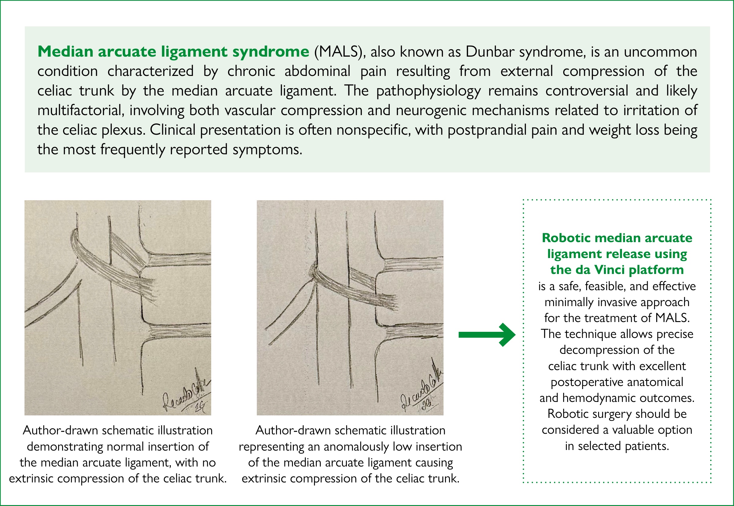
Median arcuate ligament syndrome (MALS) is an uncommon condition characterized by chronic and intermittent abdominal pain, typically postprandial, and weight loss, caused by extrinsic compression of the celiac trunk by the median arcuate ligament. The pathophysiology of the disease is not fully understood. Diagnosis is challenging due to nonspecific symptoms, and requires a careful correlation between clinical findings and imaging studies.
To demonstrate the feasibility, safety, and technical aspects of robotic median arcuate ligament release using a case-based approach with detailed imaging correlation.
The authors report the technical aspects in an elderly male patient with typical symptoms of MALS, who underwent robotic-assisted median arcuate ligament release using the da Vinci X platform. Preoperative evaluation included CT angiography and color Doppler ultrasound, demonstrating focal proximal celiac trunk stenosis.
The robotic approach allowed precise dissection and complete decompression of the celiac trunk without intraoperative complications. Postoperative imaging demonstrated resolution of the stenosis and normalization of Doppler flow parameters. The patient experienced complete symptom resolution, and remained asymptomatic after one year of follow-up.
Robotic median arcuate ligament release is a safe and effective minimally invasive option, providing excellent visualization and precise dissection in a challenging anatomical region. This technique should be considered a valuable approach for selected patients with MALS.
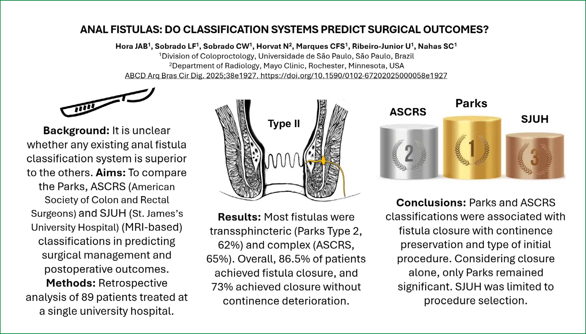
Anal fistulas remain challenging in colorectal surgery, with recurrence and postoperative incontinence common despite advances in treatment. Multiple classification systems exist, but their predictive value for surgical and functional outcomes is unclear.
The aim of this study was to compare Parks, American Society of Colon and Rectal Surgeons (ASCRS), and St. James’s University Hospital (SJUH) MRI-based classifications in predicting surgical outcomes, including continence preservation.
Retrospective analysis of 89 patients undergoing definitive surgical treatment for anorectal fistulas at a single referral center (2012–2019). Exclusions included rectovaginal fistulas, Crohn’s disease, or prior pelvic radiotherapy. Fistulas were classified using Parks, ASCRS, and, when available, SJUH (n=49). Outcomes included the number of procedures, type of initial procedure, fistula closure, and closure without continence deterioration. Continence was assessed using the Cleveland Clinic Jorge-Wexner score.
Most fistulas were transsphincteric (Parks Type 2, 62%) and complex (ASCRS, 65%). Overall, 86.5% achieved fistula closure, and 73% achieved closure without continence deterioration. Parks and ASCRS were significantly associated with fistula closure with continence preservation (p=0.008 and 0.007, respectively) and type of initial procedure. Parks remained significant when considering closure alone (p=0.005), while ASCRS showed a borderline association (p=0.051). SJUH classification was associated only with procedure selection.
Parks and ASCRS classifications were associated with fistula closure with continence preservation and type of initial procedure. Considering closure alone, only Parks remained significant. SJUH was limited to procedure selection. Overall, Parks and ASCRS guide surgical planning and prediction of functional outcomes, with Parks slightly more sensitive.
Inguinal hernia is the most frequently diagnosed hernia and affects approximately one-third of the male population. Several risk factors have been identified, including advanced age, limited physical activity, smoking, and increased intra-abdominal pressure, among others.
The aim of the study was to determine whether constipation is a risk factor for inguinal hernia in the adult population.
A case-control study was conducted at the Department of Surgery of one hospital in the north of Peru, including 121 patients with a confirmed diagnosis of inguinal hernia as cases and 242 patients without such a diagnosis as controls. Inclusion and exclusion criteria were applied, and data were collected through individual interviews using a structured questionnaire that addressed clinical aspects, lifestyles, and the presence of constipation, assessed according to the Rome IV criteria.
Results:
The results revealed significant differences between the groups of patients with and without inguinal hernia in terms of age, sex, and anthropometric characteristics. In addition, statistically significant associations were found between the presence of an inguinal hernia and type 2 diabetes, smoking, and constipation. A multivariate analysis showed that age, male sex, body mass index, high blood pressure, and constipation were significant and independent factors associated with the presence of inguinal hernia.
Constipation is a significant risk factor for inguinal hernia in the adult population. These results support the importance of considering constipation as a risk factor in the evaluation and management of patients with inguinal hernia, highlighting the relevance of adequate clinical care in this group of patients.
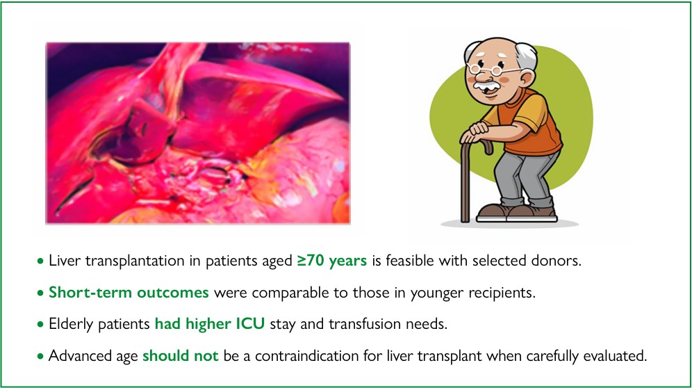
Liver transplantation (LT) is increasingly recognized as a treatment option for various diseases affecting a growing elderly population. However, its use in patients over 70 years of age remains controversial in centers with suboptimal outcomes or high waitlist mortality.
The aim of this study was to evaluate the effectiveness of LT as a treatment option for elderly patients aged 70 years or older, in comparison with younger recipients.
This retrospective study was conducted based on medical record data from 309 liver transplant recipients treated by the same surgical team across three hospitals — two located in São Paulo, São Paulo state (SP) and one in Rio Branco, Acre state (AC). Patients were divided into two groups for comparison: those aged up to 69 years (Group I) and those aged 70 years or older (Group II).
Donor characteristics were similar between the two groups, except for a higher norepinephrine dose in Group I (p<0.05). Group II showed greater transfusion requirements and longer intensive care unit (ICU) stays (p<0.05), as well as higher rates of malnutrition and comorbidities. Notably, 90-day survival was comparable between the groups.
Patients aged 70 years or older can achieve outcomes comparable to those of younger recipients, provided they receive grafts from carefully selected donors. This population should not be excluded from transplant waitlists, and specific allocation policies or scoring adjustments should be considered to ensure equitable access.
A retrospective analysis of liver transplants was performed, comparing patients over and under 70 years of age. The elderly group was transplanted with careful donor selection and obtained results comparable to those of the younger group.
This study aims to show that elderly patients over 70 years of age can have good results after liver transplantation, comparable to patients under 70 years of age, with good donor selection and perhaps additional points to favor their position on the waiting list.
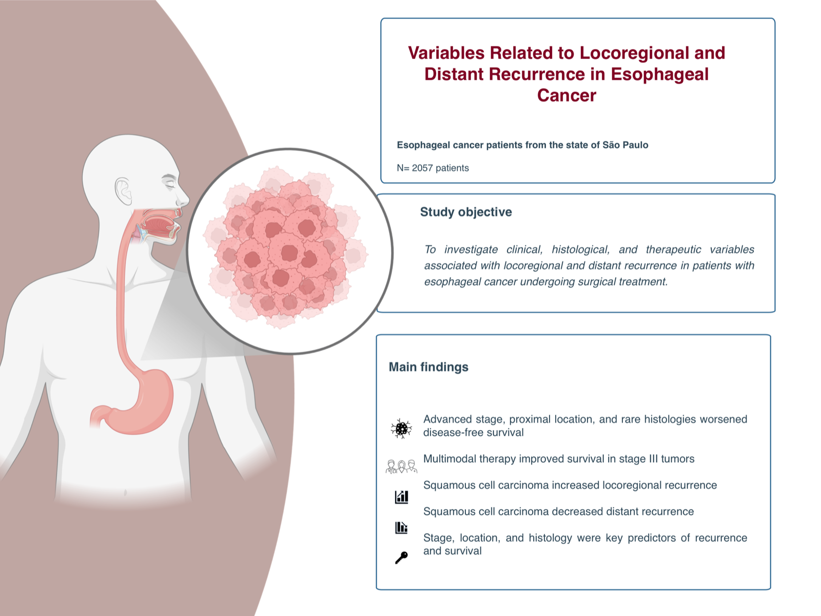
Esophageal cancer remains one of the most aggressive malignancies of the gastrointestinal tract, with high rates of recurrence and mortality despite curative-intent surgery and adjuvant therapies. Identifying factors associated with recurrence is crucial for improving outcomes and guiding personalized treatment.
The aim of this study was to evaluate pretreatment and treatment-related variables associated with recurrence in patients with esophageal cancer undergoing surgical resection.
This retrospective study analyzed data from patients with stage I–III esophageal carcinoma who underwent esophagectomy between 2000 and 2025, using the Fundação Oncocentro de São Paulo (FOSP) database. Clinical, histological, and treatment-related variables were evaluated. Disease-free survival and recurrence patterns were assessed using Cox proportional hazards models and Fine–Gray subdistribution hazard models.
A total of 2,057 patients were included, with a mean follow-up of 36.5 months (±44.8). In the multivariate analysis, advanced tumor stage (stage II: HR 1.68, 95%CI 1.21–2.33; stage III: HR 3.23, 95%CI 2.29–4.56; both p<0.01), location (middle esophagus: HR 1.31, 95%CI 1.11–1.54; p=0.001; upper esophagus: HR 1.54, 95%CI 1.21–1.96; p<0.001), and histological subtype (rare histologies: HR 2.17, 95%CI 1.35–3.49; p=0.001) were associated with worse disease-free survival. Multimodal therapy improved disease-free survival (HR 0.40, 95%CI 0.24–0.66) in stage III tumors. Squamous cell carcinoma was independently associated with locoregional recurrence (SHR 1.52, 95%CI 1.05–2.20; p=0.027). For distant recurrence, squamous cell carcinoma showed a protective effect (SHR 0.52, 95%CI 0.31–0.88; p=0.015), while high tumor grade (grade II: SHR 3.65, 95%CI 1.98–6.72; p<0.001) was associated with an increased risk. Multimodal treatments influenced recurrence patterns but did not independently predict outcomes after adjustment.
Tumor stage, location, and histology were strong predictors of disease-free survival after surgery for esophageal cancer. Histological subtypes significantly influenced recurrence patterns. Squamous cell carcinoma was associated with a higher risk of locoregional recurrence but a lower risk of distant metastasis compared to adenocarcinoma. Multimodal therapy demonstrated a protective effect in stage III disease.




Developed by Surya MKT