

Gastric cancer is the fifth most common and a leading cause of cancer death. Since 2005, perioperative chemotherapy (CT) has been the standard for non-metastatic gastric adenocarcinomas. Tumor response relies essentially on histological criteria.
The aim of the study was to evaluate tumor regression grade (TRG) after neoadjuvant CT and compare the Mandard and Becker scoring systems.
This 15-year retrospective study included patients with gastric adenocarcinoma treated with neoadjuvant CT and surgery. The TRG was assessed using Mandard and Becker scores, evaluated by area under the curve (AUC) for homogeneity, monotonicity, and discrimination. Tumors were staged by the American Joint Committee on Cancer and classified as the World Health Organization.
Forty patients (mean age 62 years; M:F ratio 2.6) were included. Tubular adenocarcinoma was the most common (48%), and 20% were stage IV. Mandard TRG1 and TRG5 each accounted for 15%, with median survivals of 48 and 30.5 months, respectively. For Becker TRG, they were 25.15 months (TRG 1), 24 months (TRG 2), and 54 months (TRG 3). The mean survival was 49.2 months for TRG1 and 39.2 months for TRG5 (Mandard), 50.3 months for TRG1 and 42.2 months for TRG3 (Becker). The positive predictive values for Mandard and Becker were 1.116 and 0.418 at 1 year and 5.719 and 1.820 at 5 years. The linearity values for Mandard and Becker were 0.6 and 0.3 at 1 year and 2.5 and 2.2 at 5 years. The AUC values at 1 year were 0.568 (Mandard), and 0.545 (Becker), and 0.606 for both at 5 years.
TRG is an independent survival predictor in gastric cancer, with similar performance between Mandard and Becker scores. Combined with ypTNM staging, it may enhance prognostic accuracy.
The analysis of tumor budding (TB) and its prognostic value in gastric adenocarcinoma (GA) has been the focus of several studies, with inconsistent results. This parameter is not included in gastric prognostic classifications or standardized pathological reports.
To evaluate TB in GA and its prognostic significance through survival analysis, in addition to investigating the association between TB and clinicopathological markers that are considered prognostic factors for this type of cancer.
This retrospective study covers a period of ten years, from January 2008 to December 2017. It included patients who underwent surgery for GA. TB evaluation followed the 2016 consensus guidelines for colorectal cancer, with three grades: Bd1 (0–4 buds), Bd2 (5–9 buds), and Bd3 (10 or more buds). Additionally, a two-grade classification system was employed, distinguishing between low-grade budding (fewer than 10 buds) and high-grade budding (10 or more buds).
TB was classified as low-grade in 69% of the cases and high-grade in 31%. High-grade TB was significantly correlated with perineural invasion (HR [hazard ratio]: 2.98, 95%CI [95% confidence interval] 1.04–8.53, p=0.004), stages III and IV (HR 4.04, 95%CI 1.27–12.83, p=0.01), and mortality (HR 3.65, 95%CI 1.24–10.74, p=0.02). It was an independent prognostic factor for recurrence-free survival (RFS) (p=0.005, p<0.05).
We have demonstrated that TB prognostic and predictive value in GA is significant, particularly regarding patient survival.
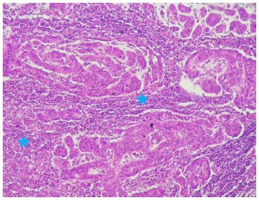
The prognostic value of tumor-infiltrating lymphocytes has been studied in several cancers, but in gastric cancer, their evaluation by standard hematoxylin-eosin staining remains controversial.
To analyze the prognostic value of tumor-infiltrating lymphocytes in gastric cancer and to investigate the association between tumor-infiltrating lymphocyte levels and a range of clinical and pathological factors in gastric cancer.
Forty-four patients were included with a mean age of 62 years, and 77% of the patients were classified as having pathological tumor stage 3 or pathological tumor stage 4. Lymph node metastases were noted in 66% of our patients. At diagnosis, 57% of patients were at an advanced stage (III). Patients with distant metastases were not included in this study. Evaluation of tumor-infiltrating lymphocytes in hematoxylin-eosin staining found that the mean tumor-infiltrating lymphocyte rate was 43 (extremes: 5-90%). 36% of patients had a tumor-infiltrating lymphocyte rate >50%. The overall survival of our patients was 40 months. Patients with tumor-infiltrating lymphocytes >50% had greater survival, without a significant correlation (p=0.275). A tumor-infiltrating lymphocyte rate =50% was correlated with age >50 years (p=0.030), presence of lymph node involvement (p=0.023), and advanced tumor stage (III+IV) (p=0.05). In a multivariate analysis, the presence of lymph node involvement (p=0.045) and advanced tumor stage (III+IV) (p=0.015) were identified as independent factors associated with a low tumor-infiltrating lymphocyte rate =50%.
Poor prognostic factors for gastric cancer (age >50 years, presence of lymph node involvement, and advanced tumor stage) were associated with a low tumor-infiltrating lymphocyte rate assessed by hematoxylin-eosin staining. However, standardization of tumor-infiltrating lymphocyte assessment is essential to allow comparability between studies and integrate this parameter into routine practice.
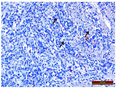
Isthmin-1, a recently discovered adipokine, was first characterized for its role in early brain development. Subsequent studies have demonstrated that isthmin-1 is involved in a broad range of biological processes, including metabolism, immunity, tumorigenesis, cellular proliferation, endothelial permeability, and organ development.
This study aimed to evaluate isthmin-1 immunoreactivity in tumor tissues obtained from gastric cancer patients using immunohistochemistry and to assess its usefulness in the diagnosis and histological evaluation of gastric cancer.
The patients were divided into three groups: adenocarcinoma, intestinal metaplasia, and healthy controls. Endoscopic biopsies, obtained according to the Sydney protocol, were examined for the control group and patients diagnosed with intestinal metaplasia. In contrast, surgical resection tissues were used for patients diagnosed with gastric adenocarcinoma.
All materials were examined histopathologically. No significant difference in isthmin-1 levels was detected between the control and intestinal metaplasia groups, based on immunohistochemical staining. However, when gastric adenocarcinoma cases were compared with the control and intestinal metaplasia groups, a significant decrease in isthmin-1 immunoreactivity was observed in adenocarcinoma cases. Patients with adenocarcinoma were grouped as well-differentiated, moderately differentiated, and poorly differentiated, and then staged according to the TNM staging system. Histopathological examination revealed no significant differences in the degree of differentiation or TNM stage.
We believe that isthmin-1 can be used as a biomarker in the diagnosis of malignant gastric diseases in the future. However, more comprehensive and extensive scientific studies are needed to determine its usefulness in tumor staging or prognosis monitoring.
Identification of epidemiological risk factors in Barrett esophagus resulting in dysplasia and adenocarcinoma and its impact on prevention and early detection.
To evaluate epidemiological risk factors involved in the development of dysplasia and esophageal adenocarcinoma from Barrett esophagus in a specific population. To critically analyze the surveillance period, aiming to individualize follow-up time according to identified risks.
A retrospective case-control study was carried out at a tertiary center involving patients diagnosed and followed up for Barrett esophagus. Patients who developed esophageal adenocarcinoma and/or dysplasia were compared to those who did not, considering variables such as gender, age, smoking status, body mass index, ethnicity, and Barrett esophagus extension. Logistic regression was performed to measure the odds ratio for risk factors associated with the outcome of adenocarcinoma and dysplasia. The presence of epidemiological risk factors in this population was correlated with the time taken to develop esophageal adenocarcinoma from metaplasia.
A statistically significant difference was observed in smoking status, race, gender, Barrett esophagus extension, and age between the group with esophageal adenocarcinoma and the group without it. Smokers and former smokers had a 4.309 times higher risk of developing esophageal adenocarcinoma, and each additional centimeter of Barrett esophagus increased the risk by 1.193 times. In the dysplasia group, smoking status, Barrett esophagus extension, and age were statistically significant factors; each additional centimeter of Barrett esophagus extension increased the risk of dysplasia by 1.128 times, and each additional year of age increased the risk by 1.023 times. Patients without risk factors did not develop esophageal adenocarcinoma within 12 months, even with prior dysplasia.
The study confirmed a higher risk of developing dysplasia and esophageal adenocarcinoma in specific epidemiological groups, allowing for more cost-effective monitorization for patients with Barrett esophagus.
Lymph node status is vital for gastric cancer (GC) prognosis, but the conventional pN stage may be limited by variations in lymphadenectomy and stage migration. The N-Ratio, which assesses the ratio of metastatic to resected lymph nodes, emerges as a promising prognostic tool.
To assess N-Ratios prognostic value in GC, particularly in patients with <25 resected lymph nodes.
Patients who underwent gastrectomy with curative intent for GC were retrospectively evaluated. The N-Ratio categories were determined using the ROC curve method, and the area under the curve (AUC) was used as a measure of performance in predicting recurrence/death.
A total of 561 GC patients were included in the study, 57% had pN+ status, and 17.5% had <25 resected lymph nodes. N-Ratio, with a mean of 0.12, predicted survival with 74% accuracy (AUC=0.74; 95%CI 0.70–0.78, p<0.001). N-Ratio categories included: N-Ratio 0 (43%); N-Ratio 1 (12.3%); N-Ratio 2 (31.6%); and N-Ratio 3 (13.2%). Disease-free survival (DFS) varied among all N-Ratio groups, with N-Ratio 3 showing worse survival than pN3 cases (DFS=21.8 vs. 11 months, p=0.022, p<0.05). In cases with <25 resected lymph nodes, DFS was not significantly worse in N-Ratio 0 (68.8 vs. 81.9%, p=0.061, p>0.05) and N-Ratio 1 (66.2 vs. 50%, p=0.504, p>0.05) groups. The DFS of N-Ratio-0 cases with <25 lymph nodes was similar to N-Ratio 1 cases.
N-Ratio influenced survival in GC patients, especially in advanced lymph node disease (N-Ratio 3). Considering that N-Ratio does not impact pN0 cases, individualized prognosis assessment is essential for patients with <25 resected lymph nodes.
Duodenal adenocarcinoma is a small percentage of gastrointestinal neoplasms, around 0.5%, and its treatment is based on resection of the tumor, classically by pancreaticoduodenectomy. In recent years, however, segmental resections of duodenal lesions, that do not involve the second portion or the periampullary region, have gained relevance with good surgical and oncological outcomes as well as the benefit of avoiding surgeries that can result in high morbidity and mortality.
To report a case of an elderly female patient with malignant neoplastic lesion in the third and fourth duodenal portion, non-obstructive, submitted to surgical treatment.
The technical option was the resection of the distal duodenum and proximal jejunum with preservation of the pancreas and reconstruction with side-to-side duodenojejunal anastomosis.
The evolution was satisfactory and the surgical margins were free of neoplasia.
Segmental resections of the duodenum are feasible and safe, offering the benefit of preventing complications of pancreaticoduodenectomies.
Gallbladder carcinoma presents a dismal prognosis. Choice treatment is surgical resection that is associated a high levels of both morbidity and mortality. Best knowledgement of prognostic factors may result a better selection of patients either for surgical or multimodal treatment.
To evaluate tecidual immunoexpression of P53, E-cadherin, Cox-2, and EGFR proteins and to correlate these findings with resected gallbladder adenocarcinoma survival.
Clinical, laboratorial, surgical, and anatomopathological reports of a series of gallbladder adenocarcinoma patients were collected by individualized questionary. Total sample was 42 patients. Median of age was 72 years (35-87). There were seven men and 35 women. Lesion distribuition in according TNM state was the following: T1 (n=2), T2 (n=5), T3 (n=31), T4 (n=4). Twenty-three patients underwent radical resection (R0), while 19 palliative surgery (R1-R2). A block of tissue microarray with neoplasic tissue of each patient was confected. It was performed evaluation of P53, E-Caderine, COX-2, and EGFR proteins imunoexpression. These findings were correlated with overall survival.
Five-year survival was 28%. The median of global survival was eight months. Only immunoexpression of EGFR protein was considered independent variable at multivariated analysis.
Final prognosis was influenced by over-expression of EGFR protein in tumoral tissue.
Gastric adenocarcinoma is more often found in men over 50 years in the form of an antral lesion. The tumor has heterogeneous histopathologic features and a poor prognosis (median survival of 15% in five years).
To estimate the relationship between the presence of nodal metastasis and other prognostic factors in sporadic gastric adenocarcinoma.
Were evaluated 164 consecutive cases of gastric adenocarcinoma previously undergone gastrectomy (partial or total), without clinical evidence of distant metastasis, and determined the following variables: topography of the lesion, tumor size, Borrmann macroscopic configuration, histological grade, early or advanced lesions, Lauren histological subtype, presence of signet ring cell, degree of invasion, perigastric lymph node status, angiolymphatic/perineural invasion, and staging.
Were found 21 early lesions (12.8%) and 143 advanced lesions (87.2%), with a predominance of lesions classified as T3 (n=99/60, 4%) and N1 (n=62/37, 8%). The nodal status was associated with depth of invasion (p<0.001) and tumor size (p<0.001). The staging was related to age (p=0.048), histological grade (p=0.003), and presence of signet ring cells (p = 0.007), angiolymphatic invasion (p = 0.001), and perineural invasion (p=0.003).
In gastric cancer, lymph node involvement, tumor size and depth of invasion are histopathological data associated with the pattern of growth/tumor spread, suggesting that a wide dissection of perigastric lymph nodes is a fundamental step in the surgical treatment of these patients.
Pancreatoduodenectomy after neoadjuvant chemotherapy is the current treatment in patients with borderline pancreatic ductal adenocarcinoma in the head of the pancreas1,2,3. The total mesopancreas excision concept includes the resection of the lymphatic structures on the right side of the SMA and along the neuronal plexus of the pancreatic head. Complete clearance of this retroperitoneal area may increase the R0 resection rate in patients with adenocarcinoma in the head of the pancreas. This area is an important location of perineural infiltration of tumor cells in patients with pancreatic ductal adenocarcinoma4.
Hackert et al5 described the term “triangle operation” as a new surgical technique for patients with locally advanced pancreatic ductal adenocarcinoma and stable disease following neoadjuvant therapy. This area is defined by SMV/PV, celiac axis/common hepatic artery, and SMA, representing the typical view after completion of the resection. However, according to the definition of the authors, the procedure should be performed without arterial resection. Recently, Loss et al6 and Schneider et al7 observed that arterial resection is effective in patients with locally advanced pancreatic cancer after neoadjuvant chemotherapy, with better long-term survival than with palliative treatment. However, this procedure should be performed in experienced pancreatic centers. After neoadjuvant chemotherapy and centers with expertise in pancreatic resection, arterial resection is perfectly possible with acceptable morbidity and mortality.




Developed by Surya MKT