

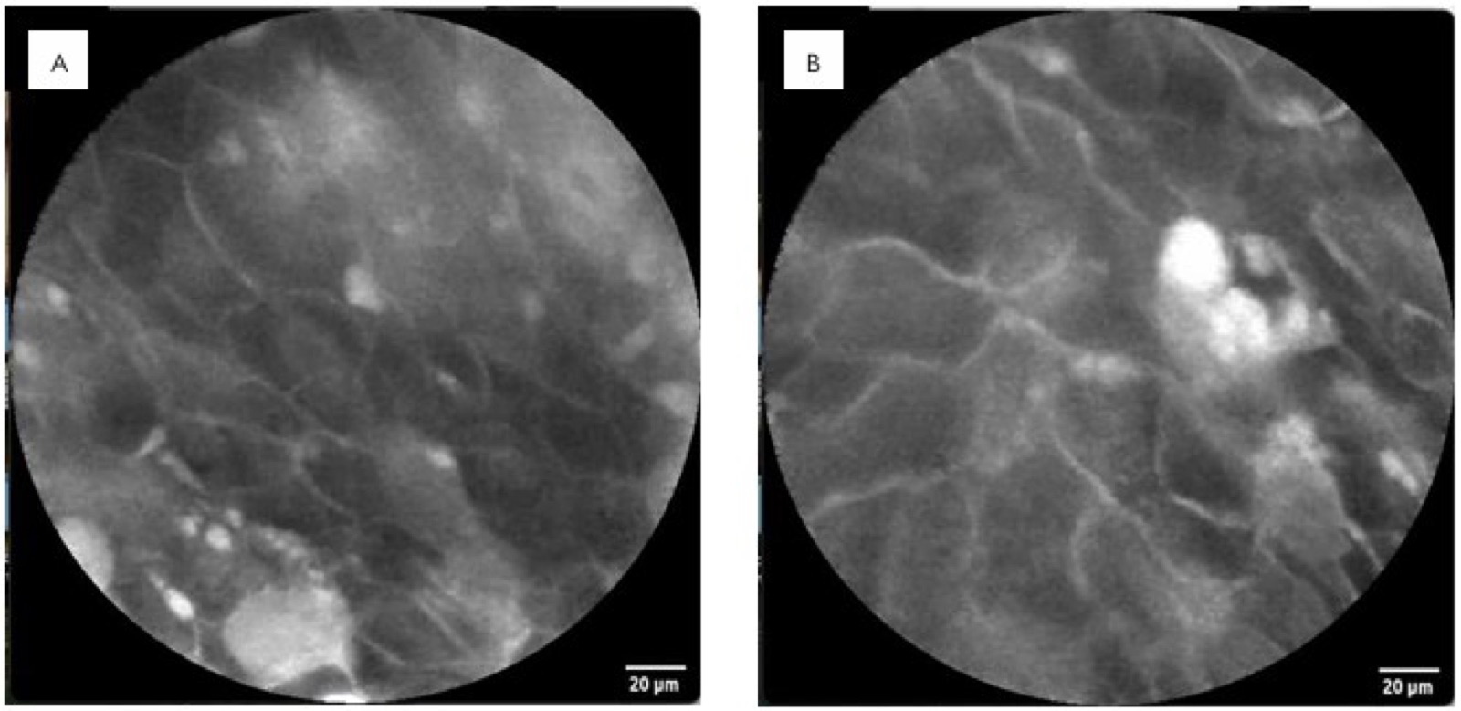
Uterus transplantation is a relatively new procedure, with successful births performed using living donors in Sweden since 2014 and a deceased donor, for the first time in Brazil, in 2016. Probe-based confocal endomicroscopy is considered an optical biopsy method (with 1000 times magnification), allowing detailed visualization of tissue cytoarchitecture and microvascular patterns at a penetration depth of approximately 50 to 60 μm. The application of confocal endomicroscopy to the uterine cervix emerges as a promising alternative to weekly cervical examinations in the follow-up of patients who have undergone uterus transplantation. The authors report the case of a 34-year-old woman with Mayer-Rokitansky-Küster-Hauser (MRKH) syndrome, diagnosed at 15 years of age, who in August 2026 underwent the first successful live-donor uterus transplantation performed in Latin America. The surgical procedure was uneventful. The confocal endomicroscopy of the uterine cervix was employed to evaluate its potential utility in identifying inflammatory changes that might precede graft rejection. No irregular or distorted epithelium or severe inflammation was observed, and this finding was confirmed by biopsies and histological analysis. They concluded that the probe-based confocal endomicroscopy may support more effective and individualized post-transplant management, representing a meaningful advancement in the fields of regenerative medicine and transplantation.




Developed by Surya MKT