

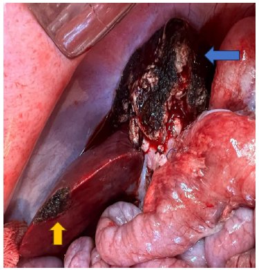
Small-for-size syndrome (SFSS) is a serious complication after partial liver transplantation or extended hepatectomy. Despite advances, the syndrome remains a complex entity with a multifactorial pathophysiology and a lack of standardized therapeutic approaches.
This study aims to establish a translational model of porcine SFSS, integrating hemodynamic, histopathological, and molecular analyses to quantify the tissue expression of biomarkers to assess liver regeneration.
Ten liver transplants were performed: eight with small (partial) for size (donors underwent a 70% hepatectomy) and two with whole liver. The recipients were followed until death (euthanasia due to severe morbidity) or for five days. Hemodynamic data, laboratory analyses, and liver tissue biopsies were collected. Immunohistochemistry was performed to assess the expression of TNF-α, IL-1β, NF-κB, and VEGF. Gene expression levels of VEGF, IL-6, IL-10, and NF-κB were quantified using reverse transcription polymerase chain reaction.
In the partial group, survival rates were 75% in the immediate postoperative period, 25% at 48 hours, and 12.5% at five days. Immunohistochemistry showed a significant increase in TNF-α and NF-κB proteins after reperfusion and at the end of surgery, respectively. Gene expression analysis showed a significant increase in IL-6 and IL-10 at the end of surgery.
This study represents a methodological advance by performing the first integrated evaluation of protein and gene expression (TNF-α, IL-6, IL-1, IL-10, VEGF, and NF-κB) correlated with hemodynamic, histopathological, and functional parameters in the small-for-size pig model.
Different lesions may affect the liver resulting in harmful stimuli. Some therapeutic procedures to treat those injuries depend on liver regeneration to increase functional capacity of this organ.
Evaluate the effects of tranexamic acid on liver regeneration after partial hepatectomy in rats.
40 rats (Rattus norvegicus albinus, Rodentia mammalia) of Wistar-UP lineage were randomly divided into two groups named control (CT) and tranexamic acid (ATX), with 20 rats in each. Both groups were subdivided, according to liver regeneration time of 32 h or seven days after the rats had been operated. The organ regeneration was evaluated through weight and histology, stained with HE and PCNA.
The average animal weight of ATX and CT 7 days groups before surgery were 411.2 g and 432.7 g, and 371.3 g and 392.9 g after the regeneration time, respectively. The average number of mitotic cells stained with HE for the ATX and CT 7 days groups were 33.7 and 32.6 mitosis, and 14.5 and 14.9 for the ATX and CT 32 h groups, respectively. When stained with proliferating cell nuclear antigen, the numbers of mitotic cells counted were 849.7 for the ATX 7 days, 301.8 for the CT 7 days groups, 814.2 for the ATX 32 hand 848.1 for the CT 32 h groups.
Tranexamic acid was effective in liver regeneration, but in longer period after partial hepatectomy.
Partial hepatectomy is a surgical intervention of the liver that can trigger its regenerative process, where the residual lobes deflagrate a compensatory hyperplasia, causing its restoration almost to the original volume. Nevertheless, depending on the extent of liver damage its regeneration might be impaired. The low-power laser has been studied with beneficial results.
To investigate the possible functional and mutagenic damage arising from the use of low-power laser used in liver regeneration after partial hepatectomy.
Fifteen male adult Wistar rats were hepatectomizated in 70% and laser irradiated or not with dose of 70 J/cm2, 650 nm, 100 mW, directly on the remaining liver, during the perioperative period. These animals were divided into four groups: G1 (control, 7 days); G2 (laser, 7 days); G3 (control, 14 days); G4 (laser, 14 days). Were analyzed the liver weight; number of hepatocytes; deposition of collagen fibers; liver function tests: serum alanine aminotransferase, aspartate aminotransferase, alkaline phosphatase, gamma glutamyl transferase, bilirubin and micronucleus test in peripheral blood erythrocyte.
The liver weight was greater in G3 and G4 (p=0.001 and p=0.002) compared to other groups. The deposition of collagen fibers in G1 was statistically higher than the other groups (p=0.01). In tests of liver function and micronucleus test was not found significant differences between the studied groups.
Low-power laser stimulation did not cause loss of liver function or mutagenic damage.




Developed by Surya MKT