

Collis gastroplasty for esophageal lengthening is a complex adjunct to hiatal hernia repair in patients with esophageal foreshortening.
To study the final morphology of the repair using state-of-the-art imaging: computed tomography with three-dimensional reconstruction.
Nine patients with prior Collis gastroplasty and hiatal hernia repair were studied with three-dimensional computed tomography reconstruction to evaluate the anatomy of the repair and screen for hiatal hernia recurrence. Secondary outcomes were quality of life and surgical morbidity.
After a medium follow-up of 34 months, objective recurrence of the hiatal hernia was observed in three patients (1.5, 2.2, and 3 cm), and two patients were symptomatic. The gastroesophageal junction tube (neo-esophagus) created by the gastroplasty was similar in shape and volume to the native esophagus in all patients. The fundoplication previously performed covered the neo-esophagus in only two of the nine patients. No fistulas or mortality were observed.
Collis gastroplasty reliably produces a tubular gastric conduit resembling the native esophagus in shape and volume. 3D CT is a promising tool for evaluating the postoperative morphology of the repair and anatomical recurrences, demonstrating the durable anatomical structure of Collis gastroplasty.
Hiatal hernias are at high risk of recurrence. Mesh reinforcement after primary approximation of the hiatal crura has been advocated to reduce this risk of recurrence, analogous to mesh repair of abdominal wall hernias. However, the results of such repairs have been mixed, at best. In addition, repairs using some type of mesh have led to significant complications, such as erosion and esophageal stricture. At present, there is no consensus as to (1) whether mesh should be used, (2) indications for use, (3) the type of mesh, and (4) in what configuration. This lack of consensus is likely secondary to the notion that recurrence occurs at the site of crural approximation. We have explored the theory that many, if not most, “recurrences” occur in the anterior and left lateral aspects of the hiatus, normally where the mesh is not placed. We theorized that “recurrence” actually represents progression of the hernia, rather than a true recurrence. This has led to our development of a new mesh configuration to enhance the tensile strength of the hiatus and counteract continued stresses from intra-abdominal pressure.
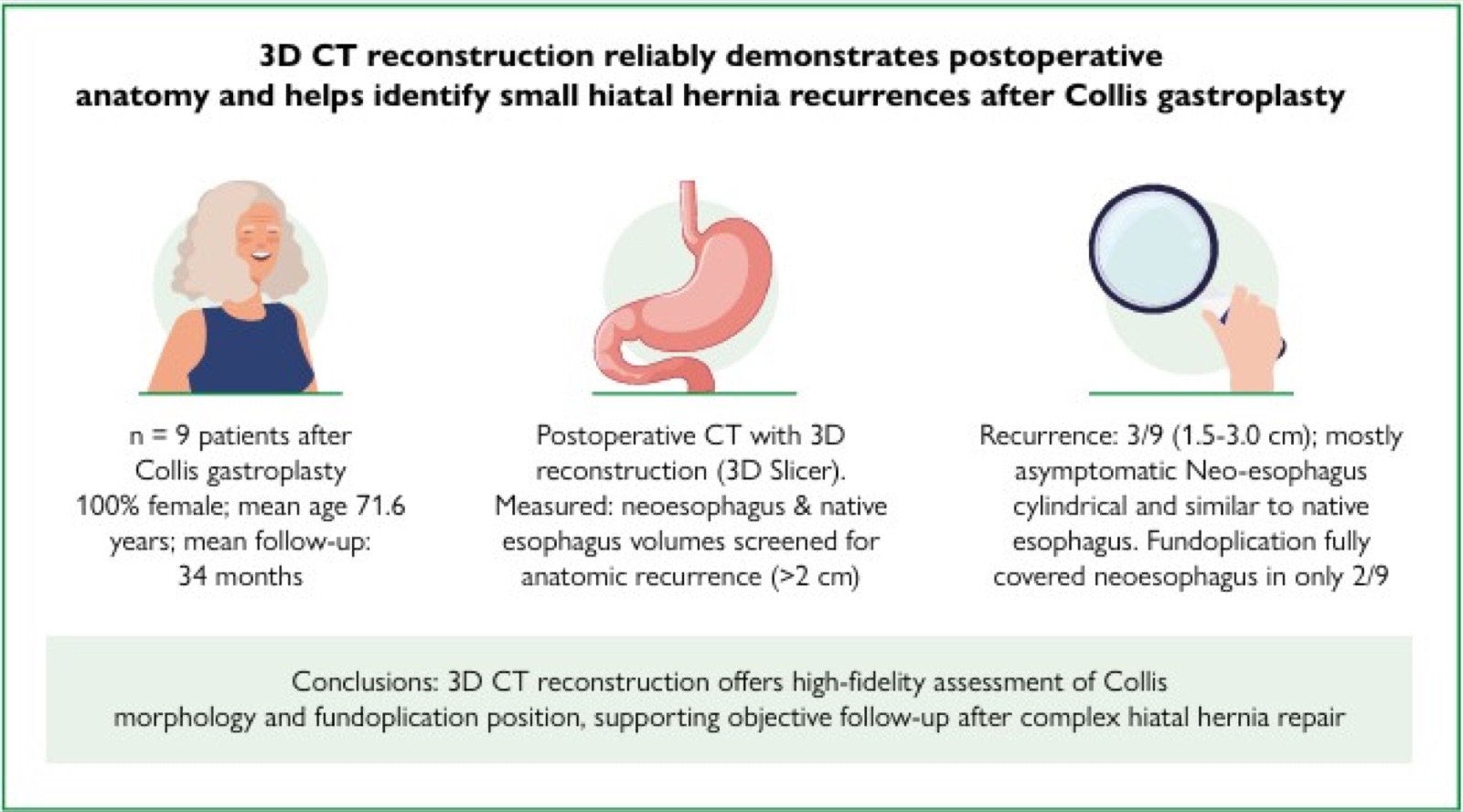
Collis gastroplasty for esophageal lengthening is a complex adjunct to hiatal hernia repair in patients with esophageal foreshortening.
To study the final morphology of the repair using state-of-the-art imaging: computed tomography with three-dimensional reconstruction.
Nine patients with prior Collis gastroplasty and hiatal hernia repair were studied with three-dimensional computed tomography reconstruction to evaluate the anatomy of the repair and screen for hiatal hernia recurrence. Secondary outcomes were quality of life and surgical morbidity.
After a medium follow-up of 34 months, objective recurrence of the hiatal hernia was observed in three patients (1.5, 2.2, and 3 cm), and two patients were symptomatic. The gastroesophageal junction tube (neo-esophagus) created by the gastroplasty was similar in shape and volume to the native esophagus in all patients. The fundoplication previously performed covered the neo-esophagus in only two of the nine patients. No fistulas or mortality were observed.
Three-dimensional computed tomography reconstruction of the gastroesophageal junction following hiatal hernia repair with Collis gastroplasty and fundoplication reliably demonstrates postoperative anatomy and helps better understand hiatal hernia recurrence.
The use of mesh in the repair of large hiatal hernias is still controversial. One of the most feared adverse events related to the use of mesh is erosion into the esophageal and gastric walls.
To record the endoscopic treatment of mesh that has migrated into the gastric lumen after surgical treatment of hiatal hernia.
The technical option was to wait for the progressive migration of the mesh into the gastric lumen, monitoring with upper digestive endoscopy, with removal by traction at the best time, with the aid of foreign body forceps.
The mesh was completely removed, and the evolution was satisfactory, without complications.
In patients with mesh migration into the stomach who are oligosymptomatic and do not show signs of complications, endoscopic surveillance and subsequent removal of the foreign body can be successfully performed when the mesh is not adhered to the gastric wall, avoiding surgical procedures with high morbidity and mortality.
The influence of body mass index on perioperative complications of hiatal hernia surgery is controversial in the surgical literature.
The aim of this study was to evaluate the influence of body mass index on perioperative complications and associated risk factors for its occurrence.
Two groups were compared on the basis of body mass index: group A with body mass index <32 kg/m2 and group B with body mass index ³32 kg/m2. A multivariate analysis was carried out to identify independent predictors for complications. Complications were classified based on the Clavien-Dindo score.
A total of 49 patients were included in this study, with 30 in group A and 19 in group B. The groups were compared based on factors, such as age, Charlson Comorbidity Index, surgical techniques used, type and location of hiatal hernia, and length of stay. Findings showed that 70% of patients had complex hiatal hernia. In addition, 14 complications also occurred: 7 pleuropulmonary and 7 requiring reoperation. From the seven reoperated, there were three recurrences, two gastrointestinal fistulas, one diaphragmatic hernia, and one incisional hernia. Complications were similar in both the groups, with type IV hiatal hernia being the only independent predictor.
Body mass index does not affect perioperative complications in anti-reflux surgery and type IV hiatal hernia is an independent predictor of its occurrence.




Developed by Surya MKT