

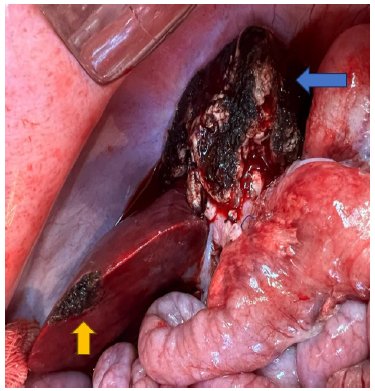
Small-for-size syndrome (SFSS) is a serious complication after partial liver transplantation or extended hepatectomy. Despite advances, the syndrome remains a complex entity with a multifactorial pathophysiology and a lack of standardized therapeutic approaches.
This study aims to establish a translational model of porcine SFSS, integrating hemodynamic, histopathological, and molecular analyses to quantify the tissue expression of biomarkers to assess liver regeneration.
Ten liver transplants were performed: eight with small (partial) for size (donors underwent a 70% hepatectomy) and two with whole liver. The recipients were followed until death (euthanasia due to severe morbidity) or for five days. Hemodynamic data, laboratory analyses, and liver tissue biopsies were collected. Immunohistochemistry was performed to assess the expression of TNF-α, IL-1β, NF-κB, and VEGF. Gene expression levels of VEGF, IL-6, IL-10, and NF-κB were quantified using reverse transcription polymerase chain reaction.
In the partial group, survival rates were 75% in the immediate postoperative period, 25% at 48 hours, and 12.5% at five days. Immunohistochemistry showed a significant increase in TNF-α and NF-κB proteins after reperfusion and at the end of surgery, respectively. Gene expression analysis showed a significant increase in IL-6 and IL-10 at the end of surgery.
This study represents a methodological advance by performing the first integrated evaluation of protein and gene expression (TNF-α, IL-6, IL-1, IL-10, VEGF, and NF-κB) correlated with hemodynamic, histopathological, and functional parameters in the small-for-size pig model.
The treatment of choice for patients with schistosomiasis with previous episode of varices is bleeding esophagogastric devascularization and splenectomy (EGDS) in association with postoperative endoscopic therapy. However, studies have shown varices recurrence especially after long-term follow-up.
To assess the impact on behavior of esophageal varices and bleeding recurrence after post-operative endoscopic treatment of patients submitted to EGDS.
Thirty-six patients submitted to EGDS were followed for more than five years. They were divided into two groups, according to the portal pressure drop, more or less than 30%, and compared with the behavior of esophageal varices and the rate of bleeding recurrence.
A significant reduction on the early and late post-operative varices caliber when compared the pre-operative data was observed despite an increase in diameter during follow-up that was controlled by endoscopic therapy.
The drop in portal pressure did not significantly influence the variation of variceal calibers when comparing pre-operative and early or late post-operative diameters. The comparison between the portal pressure drop and the rebleeding rates was also not significant.




Developed by Surya MKT